
Living with persistent knee or hip discomfort can transform simple daily tasks—like climbing stairs, taking a morning walk, or rising from a chair—into painful challenges. When initial non-surgical treatments stop providing relief, finding a path forward requires evaluating advanced surgical interventions.
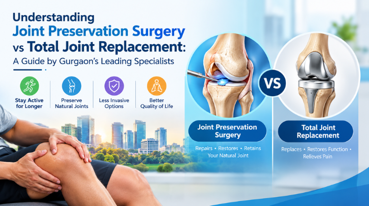
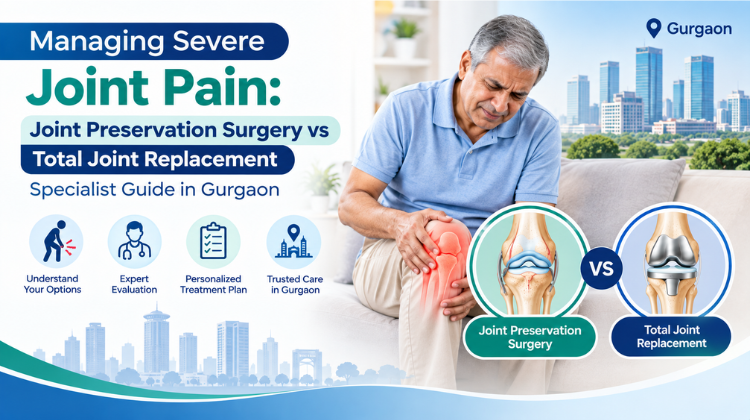
Managing severe joint pain effectively requires understanding two distinct surgical approaches: Joint Preservation Surgery and Total Joint Replacement.
Consulting an experienced orthopedic specialist in Gurgaon allows patients to navigate these options, explore modern diagnostic protocols, and select a personalized treatment plan tailored to their lifestyle, age, and joint condition.
1. Managing Severe Joint Pain: Understanding the Causes
Effective strategies for managing severe joint pain begin with identifying the structural causes of joint breakdown. Articular cartilage acts as a smooth, rubbery shock absorber protecting the ends of bones inside the joint. When this protective cushion wears away, bone-on-bone friction creates severe localized inflammation, stiffness, and pain.
┌─────────────────────────────────────────────────────────────┐
│                 Mechanisms of Joint Degeneration            │
└──────────────────────────────┬──────────────────────────────┘
                               │
       ┌───────────────────────┼───────────────────────┐
       ▼                       ▼                       ▼
┌──────────────┐       ┌───────────────┐       ┌───────────────┐
│ Post-Traumatic│      │ Mechanical    │       │ Inflammatory  │
│ Cartilage Loss│      │ Misalignment  │       │ Osteoarthritis│
└──────────────┘       └───────────────┘       └───────────────┘
The primary drivers of progressive joint destruction include:
- Osteoarthritis (OA): Progressive mechanical wear-and-tear leading to cartilage breakdown and osteophyte (bone spur) formation.
- Post-Traumatic Arthritis: Accelerated joint degradation occurring years after an acute fracture, meniscus tear, or ligament injury.
- Mechanical Malalignment: Angular deformities like bow-legs (varus) or knock-knees (valgus) that concentrate body weight onto a single compartment of the joint.
- Avascular Necrosis (AVN): Temporary or permanent loss of blood supply to the bone tissue, causing structural bone collapse (frequently observed in hip joints).
2. Managing Severe Joint Pain Through Joint Preservation Surgery
For active individuals and younger patients, managing severe joint pain does not always require installing artificial implants. Joint preservation focuses on repairing, realigning, or restoring native joint anatomy to halt degeneration and delay or prevent joint replacement.
┌─────────────────────────────────────────────────────────────┐
│             Joint Preservation Interventions                │
├─────────────────────────────────────────────────────────────┤
│ • Realignment Osteotomies (High Tibial & Distal Femoral)     │
│ • Cartilage Restoration & Repair (MACI, Microfracture)      │
│ • Arthroscopic Joint Debridement & Meniscal Repair          │
│ • Biologic Augmentations & Subchondroplasty                 │
└─────────────────────────────────────────────────────────────┘
Key Joint Preservation Procedures
1. Realignment Osteotomy (High Tibial or Femoral)
In many patients, joint arthritis is restricted to a single compartment (most commonly the inner/medial side of the knee). An osteotomy strategically reshapes the tibia or femur, shifting the body’s mechanical weight-bearing axis onto healthier, undamaged cartilage on the opposite side of the joint.
2. Cartilage Repair and Regenerative Restoration
For localized, isolated cartilage defects, specialized techniques help restore the articular surface:
- Microfracture / OATS: Creating micro-perforations in subchondral bone to stimulate blood flow and fibrocartilage growth.
- Matrix-Induced Autologous Chondrocyte Implantation (MACI): Harvesting healthy cartilage cells, culturing them on a bio-absorbable scaffold, and re-implanting them into the defect.
3. Therapeutic Arthroscopy
Minimally invasive keyhole surgery allows specialists to clear loose cartilage debris, smooth frayed joint linings, and repair torn menisci, reducing mechanical friction inside the joint.
3. Managing Severe Joint Pain with Total Joint Replacement
When joint degeneration spreads across multiple compartments or destroys subchondral bone, managing severe joint pain shifts toward joint reconstruction (arthroplasty). Total joint replacement removes damaged articular surfaces and replaces them with durable, biocompatible prosthetic components.
┌─────────────────────────────────────────────────────────────┐
│              Total Joint Replacement Components             │
└──────────────┬──────────────────────────────┬───────────────┘
               │                              │
               ▼                              ▼
┌──────────────────────────────┐┌──────────────────────────────┐
│  Prosthetic Implants         ││  Advanced Surgical Tech      │
│  • Cobalt-Chromium / Titanium││  • 3D Robotic Assistance     │
│  • Ceramic Bearings          ││  • Computer Navigation       │
│  • Polyethylene Inserts      ││  • Muscle-Sparing Approaches │
└──────────────────────────────┘└──────────────────────────────┘
Advanced Arthroplasty Approaches
- Total Knee Arthroplasty (TKA): Resurfacing the distal femur, proximal tibia, and patellar undersurface with precision-machined metal and ultra-high-molecular-weight polyethylene components.
- Total Hip Arthroplasty (THA): Replacing the damaged femoral head with a prosthetic stem and ball, paired with a smooth ceramic or metal socket cup.
- Robotic-Assisted Joint Replacement: Utilizing 3D CT imaging and robotic arms to prepare bone cuts with sub-millimeter precision, ensuring accurate implant alignment and optimal soft-tissue balance.
4. Comparing Treatment Paths: Joint Preservation vs Total Joint Replacement
Determining the appropriate path for managing severe joint pain requires comparing anatomical retention, recovery timelines, and surgical goals:
| Evaluation Criteria | Joint Preservation Surgery | Total Joint Replacement |
| Primary Objective | Preserve native bone/cartilage and shift mechanical loads | Replace worn-out joint surfaces with artificial implants |
| Anatomical Integrity | Retains natural ligaments, bone stock, and joint mechanics | Replaces articular surfaces with metal, ceramic, or polymer implants |
| Target Demographic | Younger, physically active patients (typically under 50–55) | Older adults or individuals with severe, end-stage arthritis |
| Weight-Bearing Recovery | Requires 4–8 weeks of protected weight-bearing for bone healing | Allows early weight-bearing walking within 12–24 hours post-op |
| High-Impact Activities | Often permits return to running, agility sports, and manual work | Recommends low-impact sports (walking, cycling, swimming) for implant longevity |
| Long-Term Outlook | Delays joint replacement by 10–15+ years | Modern implants offer 15–25 years of reliable joint function |
5. Specialist Assessment Criteria in Gurgaon
Consulting an orthopedic specialist in Gurgaon ensures a structured diagnostic process for managing severe joint pain.
┌─────────────────────────────────────────────────────────────┐
│              Clinical Decision Pathway                      │
└──────────────┬──────────────┬──────────────┬────────────────┘
               │              │              │
               ▼              ▼              ▼
┌──────────────────────┐┌───────────┐┌────────────────────────┐
│ Full-Length Standing ││ High-Def  ││ Patient Demographics & │
│ Alignment X-Rays     ││ MRI Scans ││ Functional Expectations│
└──────────────────────┘└───────────┘└────────────────────────┘
When evaluating treatment options, specialists review:
- Severity and Radiographic Stage: Weight-bearing X-rays assess joint space narrowing, while high-resolution MRI scans map cartilage thickness, subchondral bone health, and ligament stability.
- Mechanical Axis Realignment Potential: Full-length leg X-rays measure mechanical alignment from hip to ankle to evaluate whether an osteotomy can successfully offload damaged cartilage.
- Patient Age and Activity Goals: Younger patients who perform manual work or wish to maintain high-impact athletic routines are prioritized for joint preservation to protect native bone stock.
- Ligament Stability: Joint preservation requires intact, functional ACL/PCL or collateral ligaments. If severe instability is present, joint replacement provides a more predictable outcome.
6. Recovery and Rehabilitation Protocols
Post-operative rehabilitation plays a crucial role in long-term functional success when managing severe joint pain.
Joint Preservation Recovery Phase
- Protected Healing (Weeks 1–6): Crutches or specialized knee braces protect realigned bone cuts or cartilage repair sites while tissue heals.
- Controlled Motion: Continuous Passive Motion (CPM) devices maintain joint flexibility without applying heavy weight-bearing forces.
- Strengthening (Months 2–6): Progressive resistance training rebuilds quadriceps, hamstring, and gluteal muscle strength to support the realigned joint.
Total Joint Replacement Recovery Phase
- Early Ambulation (Day 1): Patients begin walking with support under physical therapy supervision within 24 hours of surgery.
- Functional Independence (Weeks 2–6): Focuses on restoring a natural walking gait, achieving full joint extension and flexion, and transitioning off walking aids.
- Long-Term Function: Low-impact conditioning maintains muscle tone and cardiovascular health without subjecting artificial implants to excessive wear.
#Managing Severe Joint Pain #Joint Preservation Surgery #Total Joint Replacement #Orthopedic Specialist Gurgaon #Dr Hemant Sharma #Robotic Knee Replacement #Robotic Hip Replacement #High Tibial Osteotomy #Cartilage Restoration #Total Knee Arthroplasty #Total Hip Arthroplasty #Unicompartmental Knee Replacement #FRCS Orthopedics #Joint Pain Treatment Gurgaon #Computer Navigated Surgery #Revision Joint Replacement #Adult Reconstructive Surgery #Gurgaon Orthopedic Surgeon #Sports Injury Treatment #Sub chondral Bone Repair #Ligament Sparing Surgery #Young Adult Hip Disorders #Avascular Necrosis Treatment #Minimally Invasive Joint Surgery #Knee Osteoarthritis Management #Hip Replacement Specialist #Joint Preservation Techniques #Cartilage Repair MACI
FAQ
Q1: What is the primary difference when managing severe joint pain with preservation versus replacement?
Answer:
When managing severe joint pain, joint preservation repairs, realigns, or restores your natural bones, cartilage, and intact ligaments to delay arthritis progression. In contrast, total joint replacement replaces damaged articular surfaces with biocompatible metal, ceramic, and high-density plastic implants to resolve end-stage, widespread arthritis.
Q2: Who is Dr. Hemant Sharma, and what are his primary clinical credentials in Gurgaon?
Answer:
Dr. Hemant Sharma is a prominent Orthopedic Surgeon and Chairperson- Robotic Joint Replacement & Arthroscopy Surgery, Artemis Hospital Gurugram. With over 26 years of clinical experience across India and the United Kingdom NHS system, he holds prestigious qualifications including MBBS, DNB (Ortho), MRCS (England), and FRCS (Trauma & Orthopaedics, England).
Q3: What specialized surgical techniques does Dr. Hemant Sharma use when managing severe joint pain?
Answer:
Dr. Hemant Sharma specializes in advanced robotic-assisted knee and hip replacements, computer-navigated joint surgery, complex revision arthroplasty, and joint-preserving osteotomies. He leads the Robotic Joint Replacement & Arthroscopy Surgery, Artemis Hospital Gurugram, utilizing 3D surgical planning to maximize implant accuracy, preserve muscle tissue, and speed recovery.
Q4: When should a patient choose joint preservation over total joint replacement?
Answer:
Joint preservation is recommended for active, younger patients (typically under 50–55) who have localized cartilage defects, early single-compartment wear, or angular bow-leg/knock-knee deformities. Preserving native bone delays the need for artificial implants and allows patients to maintain higher-impact physical activities.
Q5: What makes Dr. Hemant Sharma a preferred specialist for complex joint cases in Gurgaon?
Answer:
Dr. Hemant Sharma spent over 12 years receiving higher surgical training and performing complex lower limb arthroplasty in the UK NHS system. Recognized as the “Emerging Leader of the Year 2019” in orthopedics, he brings international expertise in handling high-risk joint reconstructions, complex revision surgeries, and young adult hip disorders.