Opening: 09AM - 08PM

Where Does it Hurt? Mapping High vs. Low Ankle Sprain Symptoms
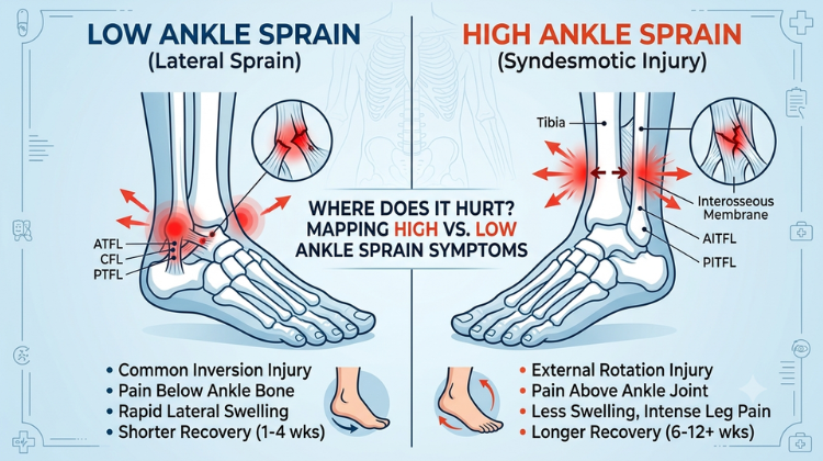
TABLE OF CONTENT

A sudden twist, an awkward landing, or a sports mishap can leave you on the ground clutching your lower leg. Ankle injuries are incredibly common, but not all sprains are created equal. When determining the right path to recovery, the most critical step is answering one key question: Where exactly does it hurt? By understanding your high vs. low ankle sprain symptoms, you can identify whether you are facing a standard lateral injury or a highly complex ligament disruption. Misdiagnosing these conditions can lead to premature weight-bearing, which significantly delays healing or causes long-term joint instability. Let’s map out the exact anatomical differences to help you decipher your discomfort.

The Physical Map: High vs. Low Ankle Sprain Symptoms Explained

Ankle sprains are broadly classified by the group of ligaments that suffer damage. Pinpointing the exact boundary of your pain tells a vivid story about what happened inside the joint.

1. Identifying Low Ankle Sprains (Lateral Inversion Injuries)

The standard or “low” ankle sprain is the type most people experience. This happens through an inversion mechanism—where your foot rolls inward underneath your ankle, stretching or tearing the lateral ligaments on the outside of the foot.

  • Pain Location: The discomfort is heavily localized on the outer edge of your ankle, primarily beneath the protruding ankle bone.

  • Visible Trait: Rapid swelling and localized bruising around the lateral side of the foot within hours.

  • Mobility: While painful, many individuals can still hobble or place partial weight on the foot shortly after the injury.

2. Spotting High Ankle Sprains (Syndesmotic Ligament Injuries)

A high ankle sprain is an entirely different clinical issue. It involves the syndesmosis—a tight network of high-tensile ligaments located above the ankle joint that binds your two lower leg bones (the tibia and fibula) securely together. This occurs when the foot is forcibly rotated outward (external rotation) or flexed upward under extreme pressure.

  • Pain Location: The pain does not sit on the outside of the foot. Instead, it radiates above the ankle joint, traveling several inches up your lower leg.

  • The “Squeeze” Indicator: If you gently squeeze your mid-calf or twist your foot outward and feel an intense, sharp pain higher up your leg, you are likely experiencing classic high vs. low ankle sprain symptoms.

  • Mobility: Bearing weight is excruciating because every step forces the tibia and fibula bones to spread apart, pulling on the torn syndesmotic tissue.

Understanding these high vs. low ankle sprain symptoms helps manage expectations, as high sprains typically take twice as long to heal compared to standard low sprains.

Clinical Diagnostic Criteria for High vs. Low Ankle Sprain Symptoms

Because the treatment paths diverge radically, medical experts utilize targeted manual testing to differentiate these injuries. When an athlete presents with swelling, a clinician will execute an external rotation test. If rotating the foot outward replicates severe pain above the joint, it points directly toward high-level ligament involvement.

Failing to properly recognize high vs. low ankle sprain symptoms early on can cause individuals to walk too soon. For a low sprain, early movement often accelerates tissue remodeling. For a high sprain, early weight-bearing continuously disrupts the healing of the syndesmosis, transforming a standard recovery into a chronic condition. 

Also Read: Is it a Normal Inversion Sprain or a High Ankle Sprain? How to Tell the Difference

When to Seek Expert Evaluation: Dr. Hemant Sharma in Gurgaon

Navigating complex ligament issues or chronic ankle pain requires a precise clinical evaluation from an expert specialist. If you are struggling to manage or differentiate your high vs. low ankle sprain symptoms, seeking top-tier medical guidance ensures an accurate recovery roadmap.

  • Current Designation: Chairman of Orthopaedics, Joint Replacement, and Spine Surgery at Marengo Asia Hospitals, Gurugram.

  • Extensive Experience: Over 31 years of rich clinical expertise spanning leading healthcare institutions across India and the United Kingdom (NHS).

  • Elite Qualifications: Holds an MBBS alongside highly prestigious certifications including the DNB (Orthopaedics), MRCS (England), and FRCS (Trauma & Orthopaedics, UK).

  • Robotic Surgery Pioneer: Recognized as a foremost first-generation robotic-assisted orthopedic surgeon in India, having successfully executed more than 500 advanced robotic procedures.

  • Acclaimed Leadership: Honored with the prestigious Service Excellence Award (2025) for Robotic Orthopaedic Surgery & Joint Replacement at the Times Network India Health Summit.

Consulting an internationally trained expert like Dr. Hemant Sharma, the best ortho surgeon in Gurgaon, provides patients with advanced diagnostics, custom rehabilitation plans, and joint preservation strategies to safeguard their long-term mobility.

Long-Term Management of High vs. Low Ankle Sprain Symptoms

Ultimately, paying attention to your body’s early warning signs protects your skeletal health. Recognizing unique high vs. low ankle sprain symptoms allows you to rest appropriately, choose the correct immobilization splint, and avoid delaying your recovery.

If your pain persists, feels deep within the lower leg, or causes an unstable sensation during movement, schedule a comprehensive structural evaluation. Entrusting your recovery to an elite medical authority like Dr. Hemant Sharma, the best ortho surgeon in Gurgaon, guarantees you receive exceptional, world-class musculoskeletal care tailored to get you safely back on your feet.

High vs. Low Ankle Sprain Symptoms: Pathomechanics, Clinical Evaluation, Diagnostic Imaging, and Surgical Management

Distinguishing between high vs. low ankle sprain symptoms is a critical diagnostic step when evaluating acute lower leg and foot trauma. While both conditions result from soft tissue micro-tearing or rupture, they affect separate anatomical structures with distinct biomechanical roles.

                                [ ACUTE ANKLE TWIST EVALUATION ]
                                                |
        +---------------------------------------+---------------------------------------+
        |                                                                               |
[ LOW ANKLE SPRAIN MECHANISM ]                                          [ HIGH ANKLE SPRAIN MECHANISM ]
        |                                                                               |
  - Plantarflexion & Inward Foot Roll                                     - Dorsiflexion & Outward Rotational Strain
  - ATFL & CFL Tissue Strain/Rupture                                      - Syndesmotic Ligament Complex Separation
  - Sub-Malleolar Lateral Pain & Local Bruising                           - Supramalleolar Shin & Mid-Calf Discomfort
                                                |
                                                v
                                  [ CLINICAL DIAGNOSTIC PATHWAY ]
                                  - Focused Stress Tests (Drawer / Squeeze / External Rotation)
                                  - Weight-Bearing Radiography & High-Res Contrast MRI
                                  - Functional Rehab vs. Rigid Boot Protection or Dynamic Suture Fixation

A low ankle sprain involves the extra-articular lateral ligament complex below the joint line, whereas a high ankle sprain disrupts the syndesmotic complex binding the tibia and fibula together above the ankle mortise. Because a high ankle sprain directly impacts the mechanical stability of the ankle socket, misidentifying a syndesmotic injury can lead to persistent joint space widening, chronic instability, and early-onset secondary osteoarthritis.

Ligamentous Architecture and Biomechanical Comparison

A clear understanding of high vs. low ankle sprain symptoms relies on recognizing the structural differences between the involved ligament networks.

+-------------------------------------------------------------------------------------------------------------------+
|                                 LIGAMENTOUS ANATOMY & BIOMECHANICAL MATRIX                                        |
+----------------------------------+---------------------------------------+----------------------------------------+
| Structural Dimension             | Low Ankle Sprain (Lateral Complex)    | High Ankle Sprain (Syndesmotic Complex)|
+----------------------------------+---------------------------------------+----------------------------------------+
| Primary Ligaments Involved       | ATFL (Anterior Talofibular Ligament), | AITFL, PITFL, Transverse Ligament, and |
|                                  | CFL (Calcaneofibular Ligament)        | Interosseous Membrane (IOM)            |
+----------------------------------+---------------------------------------+----------------------------------------+
| Primary Injury Mechanism         | Plantarflexion with inward foot roll  | External rotation of the foot with     |
|                                  | (inversion strain on lateral wall)    | forced dorsiflexion (wedging action)   |
+----------------------------------+---------------------------------------+----------------------------------------+
| Structural Function              | Limits excessive inversion and        | Binds tibia and fibula together;       |
|                                  | anterior translation of the talus     | maintains structural ankle mortise     |
+----------------------------------+---------------------------------------+----------------------------------------+
| Weight-Bearing Loading Strain    | Moderate; axial load does not push    | Severe; axial weight forces talus into |
|                                  | the outer joint walls apart           | mortise, expanding distal gap          |
+----------------------------------+---------------------------------------+----------------------------------------+

1. Lateral Extra-Articular Ligaments (Low Sprain Complex)

The lateral ligament complex stabilizes the outer side of the ankle joint below the malleolar line:

  • Anterior Talofibular Ligament (ATFL): The weakest component of the lateral complex and the first tissue disrupted during a classic inward foot twist.

  • Calcaneofibular Ligament (CFL): Connects the outer malleolus to the heel bone, limiting subtalar joint tilt.

  • Posterior Talofibular Ligament (PTFL): A strong posterior band that resists backward displacement of the talus, rarely damaged in simple twists.

These ligaments lie outside the main weight-bearing bone column. As a result, basic walking exerts minimal direct separation force on repairing lateral fibers once acute inflammation subsides.

                             [ ANKLE MORTISE & SYNDESMOSIS ARCHITECTURE ]

           Tibia (Shinbone)                                        Fibula (Outer Bone)
         +------------------+                                    +--------------------+
         |                  |====================================|                    |
         |                  |    Interosseous Membrane (IOM)     |                    |
         |                  |====================================|                    |
         |                  |      AITFL (Anterior Inferior)     |                    |
         |                  |------------------------------------|                    |
         +------------------+                                    +--------------------+
                            \                                    /
                             \--- Ankle Mortise Socket (Talus) -/

2. Syndesmotic Ligamentous Network (High Sprain Complex)

The syndesmosis acts as a high-tensile fibrous articulation connecting the distal tibia and fibula. This complex includes:

  • Anterior Inferior Tibiofibular Ligament (AITFL): Spans the front aspect of the distal tibiofibular joint.

  • Posterior Inferior Tibiofibular Ligament (PITFL) & Transverse Ligament: Heavy posterior structures providing deep structural support to the ankle socket.

  • Interosseous Membrane (IOM): A broad sheet of fibrous tissue extending up the lower leg between the tibia and fibula.

Together, these structures form a secure socket—the ankle mortise—that holds the talus bone in place. When the syndesmosis is torn, the tibia and fibula separate under weight-bearing loads, disturbing normal joint alignment.

Biomechanics of Injury: Inversion vs. External Rotation Kinematics

The anatomical distinction explains why symptoms, functional limitations, and recovery timelines vary significantly between low and high ankle sprains.

                                  [ KINEMATIC TRAUMA DYNAMICS ]
                                                |
        +---------------------------------------+---------------------------------------+
        |                                                                               |
[ INVERSION MECHANISM (LOW SPRAIN) ]                                    [ EXTERNAL ROTATION (HIGH SPRAIN) ]
        |                                                                               |
  - Ankle in plantarflexion (pointed down)                                - Ankle in dorsiflexion (flexed up)
  - Heel turns inward under body mass                                     - Foot forced outward relative to tibia
  - Tensile strain pulls across lateral ATFL/CFL                          - Wide anterior talus wedges into mortise
  - Soft tissue damage limited to sub-malleolar space                     - Forces tibia & fibula to split apart
                                                |
                                                v
                                  [ STRUCTURAL CONSEQUENCE ]
                                  - Low Sprain: Lateral ligament strain without bony widening
                                  - High Sprain: Mortise widening, high risk of latent instability

1. Plantarflexion and Inward Roll (Low Sprain Kinematics)

Standard inversion sprains occur when the foot turns inward while pointed downward (plantarflexion). This places sudden tensile strain across the ATFL and CFL. If the stress exceeds ligament capacity, micro-tearing occurs, causing pain localized entirely beneath the ankle joint line.

2. Dorsiflexion and External Rotation (High Sprain Kinematics)

A high ankle sprain occurs through a different mechanical process. When the foot is planted on the ground and flexed upward (dorsiflexion), the wider anterior section of the talus rotates back into the ankle mortise. If an outward twisting force (external rotation) is applied to the foot or lower leg, the talus acts like a wedge, pushing the tibia and fibula apart and tearing the AITFL, PITFL, and interosseous membrane.

Clinical Symptoms Mapping: High vs. Low Ankle Pain Characteristics

Comparing symptom patterns allows clinicians to evaluate whether a patient is suffering from a low ankle sprain or a syndesmotic ankle injury.

+----------------------------------------------------------------------------------------------------+
|                                    SYMPTOM MAPPING COMPARISON                                     |
+-----------------------+------------------------------------+---------------------------------------+
| Diagnostic Symptom    | Low Ankle Sprain (Lateral)         | High Ankle Sprain (Syndesmotic)       |
+-----------------------+------------------------------------+---------------------------------------+
| Primary Pain Site     | Below outer ankle bone             | Above ankle joint line, extending up  |
|                       | (sub-malleolar region)             | shin and mid-calf (supramalleolar)    |
+-----------------------+------------------------------------+---------------------------------------+
| Visible Edema         | Rapid local swelling on outer ankle| Diffuse, mild-to-moderate swelling    |
| & Ecchymosis          | within hours of injury             | higher up the lower leg               |
+-----------------------+------------------------------------+---------------------------------------+
| Squeeze Test Response | Negative (no pain provoked high)   | Positive (severe sharp pain felt in   |
|                       |                                    | upper shin when calf is compressed)   |
+-----------------------+------------------------------------+---------------------------------------+
| External Rotation     | Inward turning provokes localized  | Outward turning or flexing foot up    |
| Stress Sensitivity    | lateral outer discomfort           | provokes deep upper lower leg pain    |
+-----------------------+------------------------------------+---------------------------------------+
| Weight-Bearing Ability| Possible with limp in Grade I or   | Highly painful; stepping causes tibia |
|                       | Grade II tears                     | and fibula to spread apart under load |
+-----------------------+------------------------------------+---------------------------------------+
[ CLINICAL PRESENTATION ]
           |
           +---> Sub-Malleolar Localized Outer Discomfort  ===> Low Ankle Sprain (ATFL/CFL)
           |
           +---> Supramalleolar Pain & Positive Squeeze Test ===> High Ankle Sprain (Syndesmosis)

1. Pain Location and Visible Edema Patterns

  • Sub-Malleolar Discomfort: In a low ankle sprain, tenderness, swelling, and bruising (ecchymosis) are localized below the lateral malleolus (outer ankle bone).

  • Supramalleolar Discomfort: In a high ankle sprain, pain radiates above the ankle joint line, traveling several inches up the shinbone and mid-calf. Swelling directly over the outer ankle may appear surprisingly mild relative to the severity of internal structural damage.

2. Manual Diagnostic Stress Tests

Specialized physical stress tests help differentiate between these injuries during clinical evaluation:

  • The Squeeze Test: Compressing the mid-calf compresses the upper tibia and fibula, causing the lower ends of the bones to bow outward. Pain felt at the distal lower leg during this test indicates syndesmotic damage.

  • External Rotation Stress Test: With the knee flexed at 90 degrees, rotating the foot outward recreates sharp pain in the anterior lower leg in high ankle sprains.

  • Anterior Drawer Test: Translates the heel bone forward relative to the shinbone to assess ATFL integrity (positive in severe inversion sprains).

Advanced Diagnostic Workup: Stress Radiography, WBCT, and High-Res MRI

Accurate evaluation requires imaging protocols capable of revealing soft tissue tears and subtle joint widening under load.

[ DIAGNOSTIC IMAGING PATHWAY ]
               |
               +---> Standard AP, Lateral, and Mortise Radiographs
               |
               +---> Weight-Bearing Stress Radiography (Assess Medial & Tibiofibular Space)
               |
               +---> High-Resolution Contrast MRI (Evaluate ATFL vs. AITFL/IOM Integrity)
               |
               +---> Weight-Bearing Computed Tomography (WBCT 3D Mortise Evaluation)

1. Radiographic Evaluation and Clear Space Measurements

Plain radiographs rule out fractures, such as a distal fibular fracture or a high proximal fibular rupture (Maisonneuve fracture).

+----------------------------------------------------------------------------------------------------+
|                               RADIOGRAPHIC DIAGNOSTIC PARAMETERS                                   |
+----------------------------------+-----------------------------------------------------------------+
| Radiographic Measurement         | Diagnostic Reference Threshold                                  |
+----------------------------------+-----------------------------------------------------------------+
| Medial Clear Space (MCS)         | Normal: ≤ 4 mm. Value > 4 mm indicates talar shift and          |
|                                  | associated deltoid/syndesmotic disruption                       |
+----------------------------------+-----------------------------------------------------------------+
| Tibiofibular Clear Space (TFCS)  | Normal: < 6 mm measured 1 cm proximal to the joint line.        |
|                                  | Value ≥ 6 mm confirms syndesmotic mortise widening              |
+----------------------------------+-----------------------------------------------------------------+
| Tibiofibular Overlap (TFO)       | Normal: > 6 mm on AP view; > 1 mm on mortise view.              |
|                                  | Reduced overlap indicates tibiofibular separation               |
+----------------------------------+-----------------------------------------------------------------+

2. High-Resolution MRI and Weight-Bearing CT

  • Magnetic Resonance Imaging (MRI): The gold standard for soft tissue evaluation, MRI visualizes damaged AITFL, PITFL, and interosseous membrane fibers, allowing clinicians to grade injury severity accurately.

  • Weight-Bearing Cone-Beam CT (WBCT): Provides 3D imaging under full body weight, identifying sub-millimeter shifts or bone rotations that may not appear on standard flat radiographs.

Surgical Fixation Technologies: Rigid Screws vs. Flexible Suture-Buttons

When a high ankle sprain causes joint instability (Grade II with latent gap, or Grade III), surgical reduction and stabilization are required.

+----------------------------------------------------------------------------------------------------+
|                               SURGICAL FIXATION COMPARISON MATRIX                                  |
+----------------------------------+--------------------------------+--------------------------------+
| Surgical Parameter               | Transfixation Screws           | Dynamic Suture-Button System   |
|                                  | (3.5 mm / 4.5 mm Metallic)     | (e.g., Arthrex TightRope)      |
+----------------------------------+--------------------------------+--------------------------------+
| Primary Mechanical Behavior      | Rigid fixation;                | Dynamic semi-flexible hold;    |
|                                  | eliminates all micro-motion    | permits natural fibular motion |
+----------------------------------+--------------------------------+--------------------------------+
| Secondary Removal Surgery        | Typically required at 8–12     | Permanent implant; routine     |
| Requirement                      | weeks before full weight load  | removal step not required      |
+----------------------------------+--------------------------------+--------------------------------+
| Risk of Malreduction             | Higher risk of over-tightening | Self-centering; pulls fibula   |
|                                  | or misaligning the mortise     | into its natural tibial groove |
+----------------------------------+--------------------------------+--------------------------------+
| Postoperative Mobilization       | Delayed weight-bearing to      | Earlier progressive weight-    |
| Timeline                         | avoid screw breakage           | bearing in protective boot     |
+----------------------------------+--------------------------------+--------------------------------+
              [ RIGID TRANSFIXATION SCREW ]            [ DYNAMIC SUTURE-BUTTON SYSTEM ]
             +------------------------------+          +----------------------------------+
             | Transfixes Tibia & Fibula    |          | Multi-Strand Suture Bridging     |
             | Rigidly; Risk of Metal Fatigue|    vs    | Metallic Buttons; Preserves      |
             | Requires Removal Surgery     |          | Natural Fibular Micro-Motion     |
             +------------------------------+          +----------------------------------+

1. Traditional Rigid Transfixation Screws

Transfixation screws pass through three or four bone cortices to hold the tibia and fibula together. While effective at stabilizing the joint, rigid screws restrict the natural rotation of the fibula during walking. Patients often require a second procedure at 8 to 12 weeks to remove the screws before resuming full physical activity.

2. Flexible Dynamic Fixation (Suture-Button Technology)

Modern surgical management frequently utilizes flexible suture-button systems (such as the Arthrex TightRope). This implant uses high-strength suture material suspended between two low-profile titanium buttons placed against the outer bone walls.

  • Dynamic Joint Motion: Maintains accurate reduction of the ankle mortise while allowing natural fibular movement during walking.

  • No Routine Hardware Removal: Eliminates the need for a second surgery to remove metalwork, allowing for earlier weight-bearing and faster functional rehabilitation.

Long-Term Complications: Joint Malalignment and Post-Traumatic Osteoarthritis

Failing to properly recognize and treat high vs. low ankle sprains can lead to long-term joint complications.

                             [ UNTREATED ANKLE COMPLICATION PATHWAYS ]
                                                |
        +---------------------------------------+---------------------------------------+
        |                                                                               |
[ UNTREATED LOW SPRAIN ]                                                [ UNTREATED HIGH SPRAIN ]
        |                                                                               |
  - Chronic Lateral Ligament Laxity                                       - Persistent Tibiofibular Mortise Widening
  - Recurrent Foot "Giving Way" Episodes                                  - Asymmetric Talar Shift Inside Ankle Socket
  - Subtalar Joint Instability & Pain                                     - Joint Contact Pressure Spikes > 40%
                                                |
                                                v
                                  [ POST-TRAUMATIC OSTEOARTHRITIS ]
                                  - Progressive Chondrocyte Loss & Cartilage Erosion
                                  - Chronic Joint Pain, Stiffness, & Functional Loss
                                  - Potential Need for Ankle Fusion or Replacement

1. Chronic Lateral Ankle Instability (Low Sprain Complication)

When a severe low ankle sprain heals in a stretched position, patients can develop chronic lateral ankle instability. This results in repeated ankle twists and feelings of the joint giving way during physical activity, which may eventually require anatomical ligament reconstruction (such as the Brostrom repair).

2. Mortise Malalignment and Post-Traumatic Osteoarthritis (High Sprain Complication)

Untreated syndesmotic tears allow the ankle socket to widen. Research shows that just 1 millimeter of lateral talar shift reduces joint contact surface area by up to 42%, concentrating weight-bearing forces onto smaller cartilage regions and significantly increasing the risk of early-onset post-traumatic osteoarthritis.

Divergent Rehabilitation Protocols and Timelines

Because tissue healing demands differ significantly between low and high ankle sprains, their rehabilitation programs follow distinctly different timelines.

LOW SPRAIN RECOVERY TIMELINE:
Week 1: [ Early Mobilization ] ===> Gentle movement, balance exercises, progressive weight-bearing.
Week 2-4: [ Strengthening ]    ===> Resistance bands, single-leg balance, functional mobility.
Week 4-6: [ Full Clearance ]   ===> Return to sports and full physical activity.

HIGH SPRAIN RECOVERY TIMELINE:
Week 0-6: [ Immobilization ]   ===> Non-weight-bearing in cast/boot; absolute mortise protection.
Week 6-10: [ Partial Loading ] ===> Progressive weight-bearing, straight-plane motion exercises.
Week 10-16+: [ Agility Return] ===> Multi-directional drills, sport-specific rotational rehab.

1. Accelerated Mobilization for Low Ankle Sprains

Low sprains benefit from early functional movement. Restoring range of motion, starting light weight-bearing early, and performing balance training help speed tissue recovery and prevent joint stiffness.

2. Protected Fixation and Gradual Loading for High Ankle Sprains

Conversely, placing weight on an unhealed syndesmotic tear forces the lower leg bones apart, disrupting healing tissue. Treatment requires an initial period of strict non-weight-bearing immobilization in a boot or cast. Weight-bearing and rotational activities are introduced gradually only after clinical stability is re-established.

#Ankle Sprain Symptoms #High Ankle Sprain #Low Ankle Sprain #Syndesmosis Injury #Ankle Ligament Tear #Inversion Ankle Sprain #Ankle Pain Mapping #Ortho Surgeon Gurgaon #Dr Hemant Sharma #Marengo Asia Hospital #Best Orthopedic Doctor #Robotic Joint Surgery #Sports Injury Recovery #Ankle Rehabilitation #Ankle Joint Pain #Ligament Reconstruction #Trauma Surgeon India #FRCS Orthopaedics #Ankle Recovery Timeline #Calf Squeeze Test #External Rotation Injury #Orthopedic Care Gurugram #Joint Preservation #Ankle Swelling #Tibia And Fibula #Lateral Ligament Sprain #Chronic Ankle Instability #Foot And Ankle Specialist #Ankle Brace Recovery #Sports Medicine India #Gurgaon Orthopaedic Clinic

FAQ

Q1: How can I differentiate between high vs. low ankle sprain symptoms at home?

A: You can differentiate between them by identifying the exact location of the pain and the mechanism of injury. A low ankle sprain causes pain, bruising, and swelling on the outer side of the foot below the ankle bone, usually after the foot rolls inward. A high ankle sprain causes pain higher up the lower leg, above the ankle joint, which intensifies when you squeeze your calf muscle or rotate your foot outward.Where Does it Hurt? Mapping High vs. Low Ankle Sprain Symptoms

Q2: Why is the recovery timeline for a high ankle sprain double that of a low sprain?

A: A high ankle sprain injures the syndesmotic ligaments that hold the tibia and fibula bones together. Because your entire body weight naturally pushes these two lower leg bones apart every time you stand or take a step, the healing tissue is under constant mechanical stress. This structural weight-bearing demand causes high sprains to take twice as long to heal securely compared to a standard lateral low sprain. Where Does it Hurt? Mapping High vs. Low Ankle Sprain Symptoms

Q3: Who is the best orthopaedic surgeon in Gurgaon for complex sports and ligament injuries?

A: Dr. Hemant Sharma is widely regarded as the best orthopaedic surgeon in Gurgaon for treating complex ligament tears, sports injuries, and advanced musculoskeletal trauma. He currently serves as the Chairman of Orthopaedics at Marengo Asia Hospitals, Gurugram, and brings over 31 years of prestigious international clinical experience from both India and the UK National Health Service (NHS).Where Does it Hurt? Mapping High vs. Low Ankle Sprain Symptoms

Q4: What advanced surgical credentials make Dr. Hemant Sharma a top choice for joint care?

A: Dr. Hemant Sharma holds highly distinguished global credentials, including fellowships as a Fellow of the Royal College of Surgeons (FRCS, UK) and a Member of the Royal College of Surgeons (MRCS, England). Additionally, he is a pioneer of advanced robotic-assisted orthopedic surgery in India, with more than 500 successful robotic joint surgeries completed and a recipient of the 2025 Service Excellence Award. Where Does it Hurt? Mapping High vs. Low Ankle Sprain Symptoms

Q5: What happens if you misdiagnose high vs. low ankle sprain symptoms and walk too early?

A: Misdiagnosing a high sprain as a low sprain and walking on it prematurely can lead to chronic ankle instability, widening of the ankle mortise (the space between the lower leg bones), and long-term joint pain. While early movement can aid a low sprain, a high sprain requires prolonged immobilization to prevent the tibia and fibula from permanently separating. Where Does it Hurt? Mapping High vs. Low Ankle Sprain Symptoms
