Opening: 09AM - 08PM

Is it a Normal Inversion Sprain or a High Ankle Sprain? How to Tell the Difference

TABLE OF CONTENT

Ankle injuries are incredibly common, but not all sprains affect the same parts of your body. If you have recently twisted your ankle, you might be wondering: Is it a Normal Inversion Sprain or a High Ankle Sprain? Knowing how to tell the difference is critical, as a Normal Inversion Sprain or a High Ankle Sprain will require completely different recovery timelines, treatment plans, and rehabilitation steps.

Failing to properly identify a Normal Inversion Sprain or a High Ankle Sprain can lead to permanent joint weakness or chronic pain. Let’s dive deep into the orthopedic science behind these two distinct injuries to help you identify which one you might be dealing with.

Anatomy of the Ankle: Defining the Two Injuries

To correctly evaluate whether you have a Normal Inversion Sprain or a High Ankle Sprain, it helps to know which ligaments are under duress.

What is a Normal Inversion Sprain?

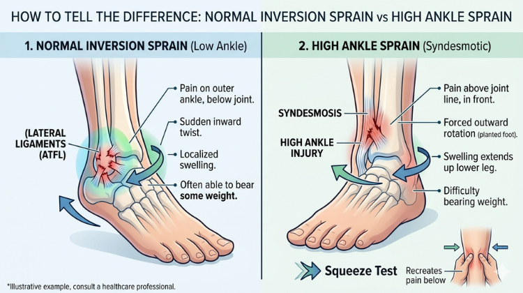
A traditional low ankle sprain happens when your foot rolls inward underneath your leg. This mechanism places extreme tension on the ligaments situated on the outside of your ankle. The most frequently damaged tissue here is the Anterior Talofibular Ligament (ATFL). Because the foot turns inward, it is anatomically referred to as an inversion trauma.

What is a High Ankle Sprain?

A high ankle sprain does not actually involve the outside of the ankle joint at all. Instead, it involves an injury to the syndesmosis. This is a complex network of tough ligaments that connects your two lower leg bones—the tibia (shinbone) and the fibula (calf bone)—above the ankle joint line. This injury occurs when your foot is planted and forced to rotate outward (external rotation) relative to your leg.

Key Symptoms: Is it a Normal Inversion Sprain or a High Ankle Sprain?

If you are struggling to categorize your injury as a Normal Inversion Sprain or a High Ankle Sprain, pay close attention to the exact location of your pain, the swelling patterns, and your ability to tolerate weight.

Diagnostic Feature Normal Inversion Sprain High Ankle Sprain (Syndesmotic)
Primary Location of Pain Outer side of the ankle, below the ankle bone. Front and outside of the lower leg, a few inches above the ankle.
Mechanism of Injury Foot rolls inward (Inversion). Foot twists outward while planted (External Rotation).
Swelling & Bruising Rapid, localized swelling around the outer ankle. Mild to moderate swelling, often spreading further up the shin.
Weight-Bearing Ability Often painful, but patients can usually limp or take a few steps. Extremely painful or completely impossible to bear weight.
The “Squeeze Test” No pain when compressing the mid-calf. Sharp pain felt at the ankle when compressing the upper calf bone.

Evaluating these clinical signs can give you a clearer indication of whether you are managing a Normal Inversion Sprain or a High Ankle Sprain.

Why Differentiating Between a Normal Inversion Sprain or a High Ankle Sprain Matters

Mismanaging a Normal Inversion Sprain or a High Ankle Sprain can have serious long-term consequences for your mobility.

If you treat a syndesmotic injury like a traditional low ankle twist, you run the risk of walking on it too early. Because a high ankle sprain destabilizes the parallel bones of your lower leg, every step you take forces the tibia and fibula to widen apart. This prevents the ligaments from knitting back together tightly.

While a minor inversion injury may feel significantly better in 2 to 4 weeks with basic rest and ice, a syndesmotic injury routinely demands 6 to 12 weeks—or potentially corrective surgery—to properly heal. Ultimately, knowing if it is a Normal Inversion Sprain or a High Ankle Sprain ensures you do not compromise your joint kinematics.

Ligamentous Architecture and Biomechanical Comparison

Evaluating the structural differences between a normal inversion sprain or a high ankle sprain requires analyzing the ligament networks that stabilize the ankle joint and lower leg.

+-------------------------------------------------------------------------------------------------------------------+
|                                  LIGAMENTOUS ANATOMY & BIOMECHANICAL MATRIX                                       |
+----------------------------------+---------------------------------------+----------------------------------------+
| Structural Dimension             | Normal Inversion Sprain (Low Sprain)  | High Ankle Sprain (Syndesmotic Injury) |
+----------------------------------+---------------------------------------+----------------------------------------+
| Anatomical Location              | Extra-articular lateral ankle aspect  | Distal tibiofibular syndesmosis        |
|                                  | below the malleolar joint line        | extending above the ankle mortise      |
+----------------------------------+---------------------------------------+----------------------------------------+
| Primary Ligaments Involved       | ATFL (Anterior Talofibular Ligament), | AITFL, PITFL, Transverse Ligament, and |
|                                  | CFL (Calcaneofibular Ligament)        | Interosseous Membrane (IOM)            |
+----------------------------------+---------------------------------------+----------------------------------------+
| Primary Injury Mechanism         | Plantarflexion with inward foot roll  | External rotation of the foot with     |
|                                  | (inversion strain on lateral wall)    | forced dorsiflexion (wedging action)   |
+----------------------------------+---------------------------------------+----------------------------------------+
| Structural Function              | Limits excessive inversion and        | Maintains structural ankle mortise;    |
|                                  | anterior translation of the talus     | prevents separation of tibia & fibula  |
+----------------------------------+---------------------------------------+----------------------------------------+
| Weight-Bearing Loading Strain    | Moderate; forces do not push the      | Severe; axial loading forces talus into|
|                                  | main lower leg bones apart            | mortise, expanding the distal gap      |
+----------------------------------+---------------------------------------+----------------------------------------+

1. Lateral Extra-Articular Ligaments (Low Sprain Complex)

In a normal inversion sprain, the pathological force is concentrated along the lateral aspect of the foot and ankle. The lateral complex comprises three primary extra-articular bands:

  • Anterior Talofibular Ligament (ATFL): The primary restrictor against anterior talar translation and internal rotation. Because it is the weakest lateral structure, it is the first tissue disrupted during an inward foot twist.

  • Calcaneofibular Ligament (CFL): Passes across the subtalar joint, stabilizing the calcaneus against excessive inversion tilt.

  • Posterior Talofibular Ligament (PTFL): A dense posterior band that resists backward displacement of the talus, rarely damaged in basic twists.

                             [ ANKLE MORTISE & SYNDESMOSIS ARCHITECTURE ]

           Tibia (Shinbone)                                        Fibula (Outer Bone)
         +------------------+                                    +--------------------+
         |                  |====================================|                    |
         |                  |    Interosseous Membrane (IOM)     |                    |
         |                  |====================================|                    |
         |                  |      AITFL (Anterior Inferior)     |                    |
         |                  |------------------------------------|                    |
         +------------------+                                    +--------------------+
                            \                                    /
                             \--- Ankle Mortise Socket (Talus) -/

2. Syndesmotic Ligamentous Network (High Sprain Complex)

Conversely, a high ankle sprain involves the fibrous articulation connecting the parallel bones of the lower leg. This network acts as a structural “zipper” and includes:

  • Anterior Inferior Tibiofibular Ligament (AITFL): Spans the front aspect of the distal tibiofibular joint.

  • Posterior Inferior Tibiofibular Ligament (PITFL) & Transverse Ligament: Heavy posterior fibrous bands forming the posterior border of the ankle socket.

  • Interosseous Membrane (IOM) and Interosseous Ligament (IOL): Broad, high-tensile fibrous sheets extending up the lower leg that transfer load between the tibia and fibula.

Biomechanics of Injury: Inversion vs. External Rotation Kinematics

Understanding the motion patterns that cause a normal inversion sprain or a high ankle sprain helps clarify why these injuries require different initial stabilization methods.

                                  [ KINEMATIC TRAUMA DYNAMICS ]
                                                |
        +---------------------------------------+---------------------------------------+
        |                                                                               |
[ INVERSION MECHANISM (LOW SPRAIN) ]                                    [ EXTERNAL ROTATION (HIGH SPRAIN) ]
        |                                                                               |
  - Ankle in plantarflexion (pointed down)                                - Ankle in dorsiflexion (flexed up)
  - Heel turns inward under body mass                                     - Foot forced outward relative to tibia
  - Tensile strain pulls across lateral ATFL/CFL                          - Wide anterior talus wedges into mortise
  - Soft tissue damage limited to sub-malleolar space                     - Forces tibia & fibula to split apart
                                                |
                                                v
                                  [ STRUCTURAL CONSEQUENCE ]
                                  - Low Sprain: Lateral ligament strain without bony widening
                                  - High Sprain: Mortise widening, high risk of latent instability

1. Plantarflexion and Inward Roll (Low Sprain Kinematics)

The vast majority of lateral sprains happen when an individual steps on an uneven surface, lands on another athlete’s foot, or turns the foot inward while the ankle is flexed downward (plantarflexion). This motion stretches the ATFL along its primary axis. If the energy exceeds tissue capacity, micro-tearing or complete rupture occurs, localized entirely beneath the ankle joint line.

2. Dorsiflexion and External Rotation (High Sprain Kinematics)

A high ankle sprain occurs through a different mechanical process. When the foot is planted firmly on the ground and flexed upward (dorsiflexion), the wider front section of the talus bone rotates into the ankle mortise. If an outward twisting force (external rotation) is applied to the foot or lower leg, the talus acts like a wedge, forcing the tibia and fibula apart and tearing the AITFL, PITFL, and interosseous membrane.

Clinical Symptoms: Identifying High vs. Low Ankle Pain

Recognizing symptom patterns allows clinicians to evaluate whether a patient has a normal inversion sprain or a high ankle sprain.

+----------------------------------------------------------------------------------------------------+
|                                    SYMPTOM MAPPING COMPARISON                                     |
+-----------------------+------------------------------------+---------------------------------------+
| Diagnostic Symptom    | Normal Inversion Sprain            | High Ankle Sprain (Syndesmosis)       |
+-----------------------+------------------------------------+---------------------------------------+
| Primary Pain Site     | Below outer ankle bone             | Above ankle joint, extending up shin  |
|                       | (sub-malleolar region)             | and mid-calf (supramalleolar region)  |
+-----------------------+------------------------------------+---------------------------------------+
| Visible Edema         | Rapid local swelling on outer ankle| Diffuse, mild-to-moderate swelling    |
| & Ecchymosis          | within hours of injury             | higher up the lower leg               |
+-----------------------+------------------------------------+---------------------------------------+
| Squeeze Test Response | Negative (no pain provoked high)   | Positive (severe sharp pain felt in   |
|                       |                                    | upper shin when calf is compressed)   |
+-----------------------+------------------------------------+---------------------------------------+
| Rotation Strain       | Inward turning of foot provokes    | Outward turning or flexing foot up    |
| Sensitivity           | localized lateral discomfort       | provokes deep lower leg pain          |
+-----------------------+------------------------------------+---------------------------------------+
| Weight-Bearing Ability| Often possible with limp in Grade I| Highly painful; stepping causes tibia |
|                       | or Grade II tears                  | and fibula to spread apart under load |
+-----------------------+------------------------------------+---------------------------------------+
[ PATIENT CLINICAL PRESENTATION ]
                |
                +---> Sub-Malleolar Pain & Localized Outer Bruising  ===> Normal Inversion Sprain
                |
                +---> Supramalleolar Pain & Positive Squeeze Test   ===> High Ankle Sprain

1. Pain Location and Visible Edema Patterns

  • Sub-Malleolar Discomfort: In a normal inversion sprain, tenderness, swelling, and ecchymosis (bruising) are heavily localized below the lateral malleolus ( outer ankle bone).

  • Supramalleolar Discomfort: In a high ankle sprain, pain radiates above the ankle joint line, traveling several inches up the shinbone and mid-calf. Localized swelling over the outer ankle may appear surprisingly minimal relative to the severity of internal structural damage.

2. Manual Physical Stress Diagnostics

Orthopedic specialists use manual provocation tests to differentiate between these injuries:

  • Anterior Drawer Test: Translates the heel bone forward relative to the shinbone to evaluate ATFL integrity (positive in severe inversion sprains).

  • Syndesmotic Squeeze Test: Compresses the mid-calf tibia and fibula together. Provoking pain down at the distal lower leg confirms a syndesmotic injury.

  • External Rotation Test: Stabilizes the lower leg while gently rotating the foot outward. Pain provoked in the lower leg indicates syndesmotic separation.

Diagnostic Workup: Radiography, Stress Imaging, and Advanced MRI

Accurate diagnosis requires advanced imaging protocols to evaluate joint alignment and soft tissue integrity.

[ DIAGNOSTIC IMAGING PATHWAY ]
               |
               +---> Standard Anteroposterior (AP), Lateral, and Mortise Radiographs
               |
               +---> Weight-Bearing Stress Radiography (Assess Medial & Tibiofibular Space)
               |
               +---> High-Resolution Contrast MRI (Evaluate ATFL vs. AITFL/IOM Integrity)
               |
               +---> Dynamic Ultrasound Assessment (Real-Time Structural Widening Assessment)

1. Standard and Weight-Bearing Radiography

Plain radiographs rule out associated fractures, such as a distal fibular fracture or a high proximal fibular rupture (Maisonneuve fracture).

+----------------------------------------------------------------------------------------------------+
|                               RADIOGRAPHIC DIAGNOSTIC PARAMETERS                                   |
+----------------------------------+-----------------------------------------------------------------+
| Radiographic Measurement         | Diagnostic Reference Threshold                                  |
+----------------------------------+-----------------------------------------------------------------+
| Medial Clear Space (MCS)         | Normal: ≤ 4 mm. Value > 4 mm indicates talar shift and          |
|                                  | associated deltoid/syndesmotic disruption                       |
+----------------------------------+-----------------------------------------------------------------+
| Tibiofibular Clear Space (TFCS)  | Normal: < 6 mm measured 1 cm proximal to the joint line.        |
|                                  | Value ≥ 6 mm confirms syndesmotic mortise widening              |
+----------------------------------+-----------------------------------------------------------------+
| Tibiofibular Overlap (TFO)       | Normal: > 6 mm on AP view; > 1 mm on mortise view.              |
|                                  | Reduced overlap indicates tibiofibular separation               |
+----------------------------------+-----------------------------------------------------------------+

2. High-Resolution Magnetic Resonance Imaging (MRI)

High-resolution MRI provides detailed visualization of damaged soft tissues. It allows clinicians to distinguish between a partial lateral band strain and a complete rupture of the AITFL, PITFL, and interosseous membrane, guiding appropriate surgical or conservative management decisions.

Surgical Management and Modern Fixation Technologies

When evaluating treatment options for a normal inversion sprain or a high ankle sprain, surgical intervention is far more common for unstable high sprains than for low sprains. While low ankle sprains rarely require acute surgery unless chronic instability develops, an unstable high sprain with mortise widening requires surgical stabilization.

+----------------------------------------------------------------------------------------------------+
|                               SURGICAL FIXATION COMPARISON MATRIX                                  |
+----------------------------------+--------------------------------+--------------------------------+
| Surgical Parameter               | Transfixation Screws           | Dynamic Suture-Button System   |
|                                  | (3.5 mm / 4.5 mm Metallic)     | (e.g., Arthrex TightRope)      |
+----------------------------------+--------------------------------+--------------------------------+
| Primary Mechanical Behavior      | Rigid rigid fixation;          | Dynamic semi-flexible hold;    |
|                                  | eliminates all micro-motion    | permits natural fibular motion |
+----------------------------------+--------------------------------+--------------------------------+
| Secondary Removal Surgery        | Typically required at 8–12     | Permanent implant; routine     |
| Requirement                      | weeks before full weight load  | removal step not required      |
+----------------------------------+--------------------------------+--------------------------------+
| Risk of Malreduction             | Higher risk of over-tightening | Self-centering; pulls fibula   |
|                                  | or misaligning the mortise     | into its natural tibial groove |
+----------------------------------+--------------------------------+--------------------------------+
| Postoperative Mobilization       | Delayed weight-bearing to      | Earlier progressive weight-    |
| Timeline                         | avoid screw breakage           | bearing in protective boot     |
+----------------------------------+--------------------------------+--------------------------------+
              [ RIGID TRANSFIXATION SCREW ]            [ DYNAMIC SUTURE-BUTTON SYSTEM ]
             +------------------------------+          +----------------------------------+
             | Transfixes Tibia & Fibula    |          | Multi-Strand Suture Bridging     |
             | Rigidly; Risk of Metal Fatigue|    vs    | Metallic Buttons; Preserves      |
             | Requires Removal Surgery     |          | Natural Fibular Micro-Motion     |
             +------------------------------+          +----------------------------------+

1. Traditional Transfixation Screw Fixation

Transfixation screws pass through three or four bone cortices to secure the tibia and fibula together. While rigid, this technique restricts natural fibular rotation during walking. Patients typically require a second procedure at 8 to 12 weeks to remove the screws before resuming high-impact activities.

2. Flexible Dynamic Fixation (Suture-Button Technology)

Modern surgical management often utilizes dynamic flexible fixation systems (such as the Arthrex TightRope). This technique uses high-strength suture material suspended between two titanium buttons placed against the outer bone walls.

  • Preserves Joint Dynamics: Allows the fibula to rotate naturally within the tibial groove during movement.

  • Eliminates Hardware Removal: Routine hardware removal is unnecessary, enabling earlier weight-bearing and faster functional rehabilitation.

Long-Term Complications: Chronic Instability vs. Mortise Malalignment

Failing to correctly diagnose a normal inversion sprain or a high ankle sprain can lead to long-term joint complications.

                             [ UNTREATED ANKLE COMPLICATION PATHWAYS ]
                                                |
        +---------------------------------------+---------------------------------------+
        |                                                                               |
[ UNTREATED INVERSION SPRAIN ]                                          [ UNTREATED HIGH SPRAIN ]
        |                                                                               |
  - Chronic Lateral Ligament Laxity                                       - Persistent Tibiofibular Mortise Widening
  - Recurrent Foot "Giving Way" Episodes                                  - Asymmetric Talar Shift Inside Ankle Socket
  - Progressive Subtalar Instability & Pain                               - Joint Stress Spikes by Over 40%
                                                |
                                                v
                                  [ END-STAGE DEGENERATIVE JOINT DISEASE ]
                                  - Progressive Chondrocyte Losses & Cartilage Erosion
                                  - Post-Traumatic Osteoarthritis & Joint Stiffness
                                  - Potential Need for Ankle Fusion or Replacement Surgery

1. Chronic Lateral Ankle Instability (Low Sprain Complication)

When a severe low ankle sprain heals in a stretched position, patients can develop chronic lateral ankle instability (CLAI). This leads to repeated ankle twisting, persistent tenderness, and feelings of the joint “giving way” on uneven ground. Chronic cases may require anatomical reconstruction using procedures like the Brostrom-Gould repair.

2. Mortise Malalignment and Post-Traumatic Osteoarthritis (High Sprain Complication)

Untreated syndesmotic tears allow the ankle socket to widen. Research shows that just 1 millimeter of lateral talar displacement reduces joint contact area by up to 42%, concentrating forces onto smaller areas of articular cartilage and significantly increasing the risk of early-onset post-traumatic osteoarthritis.

Divergent Rehabilitation Timelines and Protocols

Because tissue demands differ between a normal inversion sprain or a high ankle sprain, their rehabilitation schedules follow distinctly different timelines.

LOW SPRAIN RECOVERY TIMELINE:
Week 1: [ Early Mobilization ] ===> Gentle motion, balance exercises, progressive weight-bearing.
Week 2-4: [ Strengthening ]    ===> Resistance bands, single-leg stance, functional activities.
Week 4-6: [ Full Clearance ]   ===> Return to sports and full activity.

HIGH SPRAIN RECOVERY TIMELINE:
Week 0-6: [ Immobilization ]   ===> Non-weight-bearing in cast/boot; absolute mortise protection.
Week 6-10: [ Partial Loading ] ===> Progressive weight-bearing, straight-plane motion.
Week 10-16+: [ Agility Return] ===> Multi-directional drills, sport-specific rotational rehab.

1. Accelerated Mobilization for Low Ankle Sprains

Low sprains benefit from early, controlled movement. Restoring ankle movement, starting light weight-bearing early, and performing balance exercises speed up tissue healing and prevent long-term joint stiffness.

2. Protected Fixation and Gradual Loading for High Ankle Sprains

In contrast, early weight-bearing on an unhealed syndesmotic tear repeatedly forces the tibia and fibula apart, disrupting delicate collagen repair. Treatment requires an initial period of strict non-weight-bearing boot or cast immobilization. Weight-bearing and rotational exercises are introduced gradually only after clinical stability is re-established.

Dr. Hemant Sharma

  • Current Leadership Position: Chairman of the Department of Orthopaedics, Joint Replacement & Spine Surgery at Marengo Asia Hospitals, Gurugram.

  • Decades of Experience: Accumulates over 26 years of surgical and clinical expertise split across premier medical systems in India and England.

  • Prestigious Fellowship: Earned the elite Fellowship of the Royal College of Surgeons (FRCS) in Trauma & Orthopedic Surgery from England in 2010.

  • Specialized UK Training: Completed extensive, advanced clinical fellowships within the UK National Health Service (NHS), focusing deeply on complex sports injuries, revision surgeries, and adult reconstructive trauma.

  • Accolades & Recognition: Honored with the prestigious Service Excellence Award in Robotic Orthopaedics Surgery & Joint Replacement at the Times Network India Health Summit & Awards.

Conclusion: Consulting the Right Specialist

Determining whether you are suffering from a Normal Inversion Sprain or a High Ankle Sprain is the foundational step toward safeguarding your long-term athletic mobility. While standard sprains recover relatively quickly with conservative home care, a high ankle sprain demands precise, proactive management from an expert. If you are experiencing persistent lower leg pain, swelling above the joint line, or an inability to put weight on your foot, do not leave your recovery to chance.

For patients seeking absolute precision in care, consulting the best orthopedic surgeon in Gurgaon, such as Dr. Hemant Sharma at Marengo Asia Hospitals, ensures you receive a definitive diagnosis and an optimized rehabilitation pathway tailored to get you back on your feet safely.

#HighAnkleSprainSymptoms #LowAnkleSprain #AnkleLigamentTear #SyndesmosisInjury #AnkleInversionTrauma #ExternalRotationInjury #AnkleSqueezeTest #AnklePainAboveJoint #AnkleInstability #SportsAnkleInjury #OrthopedicAnatomy #TibiaAndFibula #ATFLLigament #AnkleSwelling #AnkleBruising #WeightBearingPain #AnkleXray #AnkleMRI #AnkleJointMechanics #LigamentHealingTime #OrthopedicSurgeonGurgaon #DrHemantSharma #MarengoAsiaHospital #SportsMedicineGurgaon #TraumaSurgery #AnkleRehabilitation #FootAndAnkleSpecialist #AnkleImmobilization #AnkleArthritisPrevention #JointPreservation

FAQ

1. Is it a normal inversion sprain or a high ankle sprain: how do doctors tell them apart?

To distinguish between a Normal Inversion Sprain or a High Ankle Sprain, an orthopedic surgeon reviews the mechanism of injury and performs localized stress exams. Doctors use the “Squeeze Test” by compressing the calf; if this recreates pain down near the ankle, it points to a high ankle sprain. They will also request weight-bearing X-rays or an MRI to visualize whether the lower leg bones have begun to widen or separate. Is it a Normal Inversion Sprain or a High Ankle Sprain? How to Tell the Difference

2. Can you walk with a high ankle sprain vs a traditional low ankle sprain?

With a traditional low ankle sprain, walking is uncomfortable but typically possible within a few days as long as the ankle is supported. However, with a high ankle injury, bearing weight pushes the ankle bones apart, causing severe, deep pain. Walking without a protective boot on an undiagnosed high ankle injury can cause long-term joint instability. Is it a Normal Inversion Sprain or a High Ankle Sprain? How to Tell the Difference

3. Who is the best doctor to consult for a complex sports ankle injury in Delhi NCR?

Dr. Hemant Sharma is widely considered an exceptional specialist for complex sports injuries, trauma, and advanced lower limb joint conditions. Serving as the Chairman of the Department of Orthopaedics, Joint Replacement & Spine Surgery at Marengo Asia Hospitals, Gurugram, he leverages more than 26 years of rigorous clinical practice across India and the United Kingdom to deliver elite musculoskeletal care. Is it a Normal Inversion Sprain or a High Ankle Sprain? How to Tell the Difference

4. How does an expert like Dr. Hemant Sharma treat a severe high ankle sprain?

For a severe high ankle injury, an expert like Dr. Hemant Sharma first assesses structural stability through advanced diagnostic imaging. If the syndesmosis is intact, conservative management utilizing a non-weight-bearing cast or customized boot is prescribed. If there is a complete ligament rupture with bone separation, he performs precise, minimally invasive stabilization surgery using syndesmotic screws or suture-button devices to restore perfect joint alignment. Is it a Normal Inversion Sprain or a High Ankle Sprain? How to Tell the Difference

5. What are the long-term risks of ignoring a high ankle ligament injury?

Ignoring a high ankle injury can cause the tibia and fibula to heal with a permanent gap between them. This structural gap alters how your foot bones rotate, leading to chronic ankle instability, a permanently reduced range of motion, recurrent micro-trauma during physical activities, and a heavily accelerated progression toward early-onset ankle arthritis. Is it a Normal Inversion Sprain or a High Ankle Sprain? How to Tell the Difference