Opening: 09AM - 08PM

Ankle sprains are among the most common orthopedic complaints worldwide, but not all sprains are created equal. If you have been diagnosed with a Syndesmotic Ankle Injury, you have likely noticed that your recovery timeline looks drastically different from a typical ankle twist. Often termed a “high ankle sprain,” this specific condition frequently leaves patients asking: Why does a Syndesmotic Ankle Injury take so long to heal?

Understanding the intricate orthopedic science behind this complex condition helps clarify why patience and specialized care are absolutely critical for a full recovery.

The Anatomy: What Makes a Syndesmotic Ankle Injury Different?

TABLE OF CONTENT

To understand why a Syndesmotic Ankle Injury is notoriously slow to heal, we must first look at the specialized anatomy of the lower leg.

A standard ankle sprain involves the lateral ligaments on the outside of the ankle. In contrast, a Syndesmotic Ankle Injury damages the syndesmosis—a complex network of ligaments (including the anterior inferior tibiofibular ligament and the interosseous membrane) that tethers your two lower leg bones together: the tibia (shinbone) and the fibula (calf bone).

This high-pressure structural network acts as a shock absorber. Every single time you take a step, stand up, or twist your foot, immense forces push these two bones apart. The primary job of the syndesmosis is to prevent this separation and maintain a rigid, stable socket (called the ankle mortise) for your foot to rotate within.

Orthopedic Science: Why a Syndesmotic Ankle Injury Delays Healing

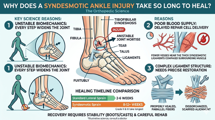
The biological and mechanical realities of the human lower body create a “perfect storm” that prolongs recovery from a Syndesmotic Ankle Injury. Orthopedic specialists highlight three primary scientific reasons for this delayed timeline:

1. Constant Mechanical Stress and Extreme Weight-Bearing Forces

Unlike ligaments in the upper body, the syndesmosis cannot easily be put at complete rest unless a patient is strictly non-weight-bearing. Every time you stand, your body weight forces the talus (the main ankle bone) upward like a wedge, naturally pushing the tibia and fibula apart. Because these injured tissues are constantly subjected to widening forces, micro-disruptions occur frequently during early healing, continuously resetting the recovery clock.

2. Poorer Vascularization and Blood Supply

Ligaments generally have a limited blood supply compared to muscles or bones, but the deep interosseous membrane and high ankle complexes are particularly poorly vascularized. Because blood carries the crucial oxygen, nutrients, and cellular building blocks required to repair torn collagen fibers, this restricted local circulation fundamentally slows down the natural cellular synthesis required to fix a Syndesmotic Ankle Injury.

3. Disruption of Joint Kinematics and Rotational Stability

High ankle sprains are almost always caused by external rotation—where the foot twists outward relative to the leg. This rotational trauma alters the exact tracking of the ankle joint. Even a tiny 1-millimeter shift or widening of the ankle mortise alters joint mechanics permanently if not healed tightly. The body requires significantly more time to re-establish this absolute rotational stability so you can safely return to twisting or pivoting actions.

Also Read:  High Ankle Sprain vs. Normal Sprain: Why the Syndesmosis Takes Twice as Long to Heal

Typical Recovery Timeline for a Syndesmotic Ankle Injury

Because of the orthopedic science outlined above, a low-grade classic sprain might resolve in 2 to 4 weeks, whereas a true Syndesmotic Ankle Injury typically requires a vastly prolonged rehabilitation track.

  • Grade I (Mild Micro-tears): 6 to 8 weeks of protected immobilization and targeted physical therapy.

  • Grade II (Partial Tears): 8 to 12+ weeks, often requiring specialized boots or customized orthotics to prevent the tibia and fibula from separating.

  • Grade III (Complete Rupture/Instability): May require surgical stabilization (such as syndesmotic screws or flexible suture-button fixations) followed by 4 to 6 months of strict, progressive rehabilitation.

Rushing back too early from a Syndesmotic Ankle Injury without restoring full structural integrity frequently leads to chronic ankle instability, persistent pain, and early-onset localized arthritis.

Detailed Structural Anatomy and Pathomechanics of the Distal Tibiofibular Joint

Understanding the mechanical behavior of a syndesmotic ankle injury requires evaluating the specific connective tissues that secure the ankle mortise.

+-------------------------------------------------------------------------------------------------------------------+
|                                 SYNDESMOTIC ANATOMY & FUNCTIONAL MATRIX                                           |
+----------------------------------+---------------------------------------+----------------------------------------+
| Structural Component             | Anatomic Location & Composition       | Primary Biomechanical Role             |
+----------------------------------+---------------------------------------+----------------------------------------+
| Anterior Inferior Tibiofibular   | Anterolateral joint line spanning     | Primary barrier against external       |
| Ligament (AITFL)                 | distal tibia to fibula                | rotation of the fibula                 |
+----------------------------------+---------------------------------------+----------------------------------------+
| Posterior Inferior Tibiofibular  | Deep posterolateral border; thicker  | Resists posterior fibular translation   |
| Ligament (PITFL)                 | and stronger than AITFL               | and backward talar displacement        |
+----------------------------------+---------------------------------------+----------------------------------------+
| Transverse Tibiofibular          | Deep inferior portion of PITFL;       | Forms posterior labrum of ankle socket;|
| Ligament                         | fibrocartilaginous tissue structure    | prevents posterior talar dislocation   |
+----------------------------------+---------------------------------------+----------------------------------------+
| Interosseous Membrane (IOM) &    | Extensive sheet-like structure running| Acts as continuous shock absorber;     |
| Interosseous Ligament (IOL)      | up the lower leg between shafts       | transfers load from tibia to fibula    |
+----------------------------------+---------------------------------------+----------------------------------------+

1. Functional Load Distribution and the Tibiofibular Vault

The distal tibiofibular joint acts as a dynamic shock-absorbing system. During normal movement, the fibula carries 10% to 15% of the body’s axial load while rotating externally up to 5 degrees during full ankle flexion. The interosseous membrane (IOM) distributes these forces across the lower leg, preventing excessive bone separation during high-impact landing, pivoting, or sprinting.

                             [ ANKLE MORTISE CROSS-SECTION ]

             Tibia (Shinbone)                                 Fibula (Calf Bone)
          +-------------------+                             +-------------------+
          |                   |=============================|                   |
          |                   |  Interosseous Membrane      |                   |
          |                   |=============================|                   |
          |                   |   AITFL / PITFL Ligaments   |                   |
          |                   |-----------------------------|                   |
          +-------------------+                             +-------------------+
                             \                               /
                              \--- Ankle Mortise Socket ----/
                                         |
                                    [  Talus  ]

2. Mechanism of Disruption: Talar Rotation Dynamics

The most common cause of a syndesmotic ankle injury is forced external rotation of the foot combined with upward bending (dorsiflexion). As the foot twists outward relative to the leg, the wider anterior section of the talus drives into the ankle mortise. This wedging force sequentially tears the anterior ligaments, the interosseous membrane, and eventually the posterior ligament structures.

Associated Traumatic Lesions: Maisonneuve Fractures and Deltoid Ligament Tears

Syndesmotic tears frequently co-occur with broader structural disruptions throughout the ankle joint and lower leg.

+----------------------------------------------------------------------------------------------------+
|                                ASSOCIATED SYNDESMOTIC TRAUMA PATTERNS                              |
+-----------------------+------------------------------------+---------------------------------------+
| Associated Injury     | Mechanical Structural Pattern      | Diagnostic Clinical Significance      |
+-----------------------+------------------------------------+---------------------------------------+
| Maisonneuve Fracture  | Complete syndesmotic rupture with  | Proximal calf tenderness requires     |
|                       | high proximal fibular fracture     | full-length tibia-fibula X-rays       |
+-----------------------+------------------------------------+---------------------------------------+
| Deltoid Ligament Complex| Medial ankle disruption occurring | Widens medial clear space (> 4 mm);   |
| Tear                  | alongside syndesmotic separation   | allows lateral shift of the talus     |
+-----------------------+------------------------------------+---------------------------------------+
| Tillaux-Chaput & Wagstaffe| Avulsion fractures of the anterior| Small bone fragments detached at      |
| Fractures             | tibial tubercle or fibular attachment| AITFL insertion points               |
+-----------------------+------------------------------------+---------------------------------------+
                                  [ MAISONNEUVE INJURY CASCADE ]
                                                |
   [ Forced External Rotation ] ===> [ Deltoid / Medial Tear ] ===> [ Syndesmosis Separation ] ===> [ Proximal Fibula Fracture ]

1. Maisonneuve Fracture Complex

A Maisonneuve fracture occurs when rotational energy travels up the lower leg, tearing the distal syndesmosis and interosseous membrane before exiting through a fracture in the upper calf bone (proximal fibular neck). Evaluating a suspected high sprain requires palpating the entire calf and obtaining full-length leg X-rays to avoid missing this high-level injury.

2. Medial Deltoid Ligament Rupture

When external rotation forces tear the syndesmotic complex, energy can continue across the inner ankle, disrupting the deltoid ligament. This combined disruption creates complete ankle instability, allowing the talus to shift outward and forward within the joint socket.

Diagnostic Workup: Stress Imaging, Dynamic Ultrasound, and Arthroscopy

Identifying subtle or latent syndesmotic instability requires specialized imaging protocols beyond standard resting X-rays.

[ SUSPECTED SYNDESMOTIC TRAUMA ]
                |
                +---> Weight-Bearing Radiographs & External Rotation Stress Views
                |
                +---> Dynamic High-Resolution Musculoskeletal Ultrasound
                |
                +---> Weight-Bearing Bilateral Computed Tomography (WBCT)
                |
                +---> Diagnostic Ankle Arthroscopy (Intraoperative Hook & Cotton Tests)

1. Stress Radiography and Weight-Bearing CT

  • Gravity and External Rotation Stress X-Rays: Applying outward pressure to the foot during imaging exposes hidden ligament gaps. A medial clear space over 4 mm or a tibiofibular clear space over 6 mm confirms structural instability.

  • Weight-Bearing Cone-Beam CT (WBCT): Provides 3D imaging under full body weight, identifying subtle bone rotations or shifts that may not appear on standard flat radiographs.

+----------------------------------------------------------------------------------------------------+
|                                DIAGNOSTIC IMAGING ACCURACY MATRIX                                 |
+----------------------------------+--------------------------------+--------------------------------+
| Imaging Modality                 | Diagnostic Capability          | Diagnostic Limitations         |
+----------------------------------+--------------------------------+--------------------------------+
| Non-Weight-Bearing Radiographs   | Identifies frank dislocations  | Misses up to 40% of latent     |
|                                  | and bone fractures             | syndesmotic instability        |
+----------------------------------+--------------------------------+--------------------------------+
| Weight-Bearing CT Scan (WBCT)    | High 3D structural accuracy;   | Requires specialized scanning  |
|                                  | detects sub-millimeter shifts  | equipment                      |
+----------------------------------+--------------------------------+--------------------------------+
| High-Res Diagnostic MRI          | Excellent visualization of soft | Non-weight-bearing; does not   |
|                                  | tissue tears (AITFL/PITFL)     | measure dynamic instability    |
+----------------------------------+--------------------------------+--------------------------------+
| Diagnostic Ankle Arthroscopy     | Direct visual inspection and   | Invasive surgical procedure    |
|                                  | mechanical probe stress testing| requiring anesthesia           |
+----------------------------------+--------------------------------+--------------------------------+

2. Intraoperative Diagnostic Ankle Arthroscopy

Diagnostic ankle arthroscopy remains the ultimate gold standard for evaluating ankle joint stability. Inserting a tiny camera into the ankle allows the surgeon to directly inspect the joint space while applying mechanical tension using a probe (the “Hook test”). Visualizing more than 2 mm of separation between the tibia and fibula confirms the need for surgical stabilization.

Surgical Fixation Strategies: Rigid Transfixation vs. Dynamic Suture-Button

When a syndesmotic ankle injury causes structural joint instability (Grade II with latent gap, or Grade III), surgical reduction and fixation are required.

+----------------------------------------------------------------------------------------------------+
|                               SURGICAL STABILIZATION COMPARISON                                   |
+----------------------------------+--------------------------------+--------------------------------+
| Surgical Technique               | Rigid Transfixation Screws     | Dynamic Suture-Button System   |
|                                  | (3.5 mm / 4.5 mm Metallic)     | (e.g., Arthrex TightRope)      |
+----------------------------------+--------------------------------+--------------------------------+
| Biomechanical Motion             | Completely rigid; eliminates   | Semi-flexible; permits normal  |
|                                  | physiological micro-motion     | physiological fibular rotation |
+----------------------------------+--------------------------------+--------------------------------+
| Hardware Removal Requirement     | Frequently requires removal at | Permanent implant; routine     |
|                                  | 8–12 weeks before full load    | removal step not required      |
+----------------------------------+--------------------------------+--------------------------------+
| Risk of Malreduction             | Higher risk of over-tightening | Self-centering; allows natural |
|                                  | the ankle mortise socket       | anatomical alignment           |
+----------------------------------+--------------------------------+--------------------------------+
| Postoperative Mobilization       | Delayed weight-bearing to      | Earlier progressive weight-   |
| Timeline                         | avoid screw fatigue or failure | bearing in protective boot     |
+----------------------------------+--------------------------------+--------------------------------+
               [ RIGID TRANSFIXATION SCREW ]            [ DYNAMIC SUTURE-BUTTON SYSTEM ]
              +------------------------------+         +----------------------------------+
              | Transfixes Tibia & Fibula    |         | Multi-Strand Suture Bridging     |
              | Rigidly; Risk of Metal Fatigue|   vs    | Metallic Buttons; Preserves      |
              | Requires Removal Procedure   |         | Natural Fibular Micro-Motion     |
              +------------------------------+         +----------------------------------+

1. Traditional Transfixation Screws

Metal syndesmotic screws pass through three or four bone walls (cortices) to hold the tibia and fibula together. While effective at stabilizing the joint, rigid screws prevent the fibula from moving naturally during walking. Because of this, screws often require a second minor surgery at 8 to 12 weeks to remove the hardware before resuming heavy physical activity.

2. Dynamic Suture-Button Fixation (TightRope Technology)

Modern surgical management increasingly utilizes dynamic flexible fixation systems, such as the Arthrex TightRope. This technique uses a strong fiber-wire suture suspended between two low-profile titanium buttons placed against the outer bone walls.

  • Preserved Joint Motion: Allows the fibula to move naturally within the tibial groove during foot flexion, lowering the risk of joint stiffness.

  • Self-Centering Alignment: Helps pull the fibula back into its natural anatomical position without over-tightening the joint.

  • No Second Removal Surgery: Eliminates the need for a second procedure to remove metalwork, allowing for earlier weight-bearing and an accelerated rehabilitation path.

Chondral Damage, Joint Kinematics, and Post-Traumatic Osteoarthritis

Untreated syndesmotic instability alters how weight is distributed across the ankle joint.

                             [ PATHOGENESIS OF POST-TRAUMATIC ARTHROSIS ]
                                                  |
        +-----------------------------------------+-----------------------------------------+
        |                                                                                   |
[ UNCORRECTED TIBIOFIBULAR GAP ]                                        [ ASYMMETRIC CARTILAGE SHEAR ]
        |                                                                                   |
  - Persistent 1 mm to 2 mm Lateral Talar Shift                          - Peak Joint Stress Spikes by > 40%
  - Reduced Contact Surface Area Across Articular Surface                - Micro-Cracking of Subchondral Bone Matrix
  - Accelerated Chondrocyte Apoptosis & Degradation                      - Progressive Joint Space Narrowing & Pain
                                                  |
                                                  v
                                    [ END-STAGE ANKLE OSTEOARTHRITIS ]
                                    - Severe Chronic Pain & Functional Limitation
                                    - Joint Stiffness & Structural Osteophyte Formation
                                    - Requires Ankle Fusion or Total Ankle Replacement

Research indicates that even a 1-millimeter lateral displacement of the talus reduces joint contact surface area by up to 42%. This shift concentrates weight onto smaller areas of articular cartilage, accelerating wear and increasing the long-term risk of post-traumatic ankle osteoarthritis.

Evidence-Based Postoperative Rehabilitation Protocols

Rehabilitation following surgical repair or conservative management of a syndesmotic ankle injury requires protecting healing tissues while progressively rebuilding joint function.

Phase 1 (Weeks 0–3):  [ Rigid Protection ]  ===> Non-weight-bearing in cast/boot; swelling management.
Phase 2 (Weeks 4–6):  [ Partial Loading ]   ===> Partial weight-bearing; straight-plane movement.
Phase 3 (Weeks 7–11): [ Dynamic Balance ]   ===> Full weight-bearing; strength & balance training.
Phase 4 (Weeks 12+):  [ Sports Return ]      ===> Multi-directional agility drills; sports clearance.

1. Phase I: Acute Protection and Swelling Management (Weeks 0–3)

Focuses on controlling inflammation and protecting healing tissue using a non-weight-bearing splint or walking boot. Active movement is limited to straight upward and downward flexing (sagittal plane), avoiding all rotational movements.

2. Phase II: Progressive Weight-Bearing (Weeks 4–6)

Gradually introduces weight-bearing while using a protective boot. Patients begin light resistance exercises with therapy bands, calf stretches, and basic balance work while avoiding twisting or pivoting motions.

3. Phase III: Proprioceptive Retraining and Dynamic Strength (Weeks 7–11)

Transitions patients into normal supportive footwear. Exercises focus on rebuilding calf strength, single-leg balance on unstable surfaces, and restoring natural gait patterns.

4. Phase IV: Sport-Specific Agility and Pivoting (Weeks 12+)

Introduces multi-directional agility drills, lateral cutting maneuvers, and dynamic jumping drills once equal leg strength and pain-free movement are achieved.

 Facts about Dr. Hemant Sharma

  • Current Designation: Chairman of the Department of Orthopaedics, Joint Replacement & Spine Surgery at Marengo Asia Hospitals, Gurugram.

  • Clinical Experience: Over 26 years of extensive medical and surgical practice across premier institutions in India and England.

  • Key Credentials: Awarded the highly prestigious Fellowship of the Royal College of Surgeons (FRCS) in Trauma & Orthopedic Surgery from England in 2010.

  • Global Training: Completed formal advanced clinical fellowships in the UK, specializing in revision procedures, complex trauma management, and complex foot and ankle configurations.

  • Specialty Focus: Highly proficient in robotic hip and knee surgeries, complex adult reconstructive trauma, joint preservation, and advanced sports injury management.

Expert Syndesmotic Reconstruction Care with Dr. Hemant Sharma

Managing complex ankle and lower leg injuries requires precise surgical evaluation, modern fixation techniques, and dedicated rehabilitation planning. Dr. Hemant Sharma is a renowned orthopedic surgeon with over 26 years of international experience in complex trauma, lower limb reconstruction, dynamic joint stabilization, and sports injury care.

+----------------------------------------------------------------------------------------------------+
|                                  DR. HEMANT SHARMA CLINICAL PROFILE                                |
+------------------------------------+---------------------------------------------------------------+
| Clinical Leadership                | Chairman – Department of Orthopedics & Joint Replacement at   |
|                                    | Marengo Asia Hospitals, Gurugram, India.                   |
+------------------------------------+---------------------------------------------------------------+
| UK NHS Fellowship Training         | Certified Fellow of the Royal College of Surgeons (FRCS,      |
|                                    | England) with advanced lower limb trauma training.           |
+------------------------------------+---------------------------------------------------------------+
| Core Orthopedic Specializations    | Syndesmotic stabilization, dynamic suture-button fixation,    |
|                                    | complex trauma reconstruction, and sports injury care.   |
+------------------------------------+---------------------------------------------------------------+

Dr. Hemant Sharma utilizes advanced diagnostic modalities, including stress radiography and high-resolution imaging, to evaluate syndesmotic injuries accurately. Whether using conservative immobilization protocols or modern dynamic suture-button fixation, Dr. Sharma helps patients achieve optimal joint stability, painless movement, and a safe return to active lifestyles.

If you are experiencing persistent lower-leg pain, ankle instability, or recovering from a rotational ankle injury, schedule a consultation with Dr. Hemant Sharma for expert evaluation and targeted orthopedic care.

#HighAnkleSprain #AnkleLigamentTear #AnkleMortiseStability #TibiofibularLigament #InterosseousMembrane #AnkleInstability #OrthopedicScience #AnkleSprainRecovery #LowerLimbTrauma #SportsInjuryRehab #AnkleJointMechanics #WeightBearingPain #LigamentHealingTime #SyndesmosisSprain #AnkleMRI #SqueezeTestAnkle #ChronicAnklePain #AnkleArthritisPrevention #AnkleImmobilization #OrthopedicSurgeonGurugram #DrHemantSharma #MarengoAsiaHospital #RoboticOrthopedicSurgery #JointPreservation #SportsMedicine #TraumaSurgery #AnkleRehabilitation #FootAndAnkleSpecialist #AnkleScrews #HighAnkleLigament #AnkleTwistHealing


FAQ

1. Why does a high ankle sprain or syndesmotic ankle injury take months to heal?

A Syndesmotic Ankle Injury takes months to heal because the affected ligaments connect the tibia and fibula bones. Every time you stand or walk, your body weight acts as a wedge that naturally forces these bones apart, placing constant stress on the healing tissues. Additionally, these deep high-ankle ligaments have a naturally poor blood supply, which severely limits the speed of cellular repair.

2. Can you walk on a syndesmotic ankle injury without surgery?

Walking immediately on a moderate-to-severe Syndesmotic Ankle Injury is highly discouraged as it widens the joint space and stalls healing. However, mild (Grade I) injuries can often heal without surgery using conservative management. This requires strict initial immobilization in a protective boot or cast, followed by a highly structured physical therapy regimen focused on rotational stability.

3. Who is the best orthopedic surgeon for complex ankle and sports injuries in Gurugram?

Dr. Hemant Sharma is widely recognized as a premier orthopedic specialist in Gurgoan for complex musculoskeletal issues, trauma, and severe joint injuries. Currently practicing as the Chairman of the Department of Orthopaedics and Joint Replacement at Marengo Asia Hospitals, Gurugram, he brings over 26 years of advanced surgical experience across India and the United Kingdom to handle intricate lower-limb conditions.

4. How does an orthopedic expert like Dr. Hemant Sharma diagnose a syndesmotic ankle injury?

An elite expert like Dr. Hemant Sharma utilizes a comprehensive clinical approach to diagnose a Syndesmotic Ankle Injury. This includes specialized physical examinations such as the “Squeeze Test” (compressing the calf to see if it reproduces ankle pain) and external rotation stress testing. This is paired with advanced diagnostic imaging, using weight-bearing X-rays to check for joint widening and high-resolution MRI to visualize deep ligament tears.

5. What happens if a severe syndesmotic ankle injury is left untreated?

If a severe Syndesmotic Ankle Injury is ignored or improperly treated, it leads to a chronic widening of the ankle mortise socket. This permanent structural instability alters normal walking patterns, causes chronic localized pain, creates a recurring sensation of the ankle “giving out,” and significantly accelerates the wear and tear of joint cartilage, leading to early-onset ankle arthritis.