Dr. Hemant Sharma
FRCS (England) MRCS (England) DNB
Specialist in Revision Lower Limb ArthroPlasty
Complex Hip, Knee and Sports Injury

Revision Knee Surgery is an excellent, life-changing procedure that offers lasting pain relief and mobility to millions of people worldwide. However, no artificial joint lasts forever. Over time, implants can wear out, become loose, or experience complications. When a primary implant fails, a second procedure known as revision knee surgery becomes necessary.
Unlike a first-time knee replacement, a secondary operation is not a straightforward routine procedure. It is a highly intricate intervention that demands specialized technical precision. Understanding the complex nature of a revision knee surgery highlights exactly why you must place your health in the hands of a seasoned orthopedic expert.
The first question many patients ask is: “Why can’t my second surgery be just like my first one?” The reality is that the anatomical environment of the knee changes dramatically after a primary replacement has been altered or removed.
Here are the key factors that contribute to the challenges of a revision knee surgery:
When an old prosthetic implant is removed, it often takes bits of the patient’s natural bone along with it. Dealing with degraded or missing bone structures means the surgeon must use specialized bone grafts, metallic augments, or long-stemmed implants to rebuild a stable foundation for the new joint.
The original surgery leaves behind dense scar tissue. Accessing the knee joint during a revision knee surgery requires navigating through this altered tissue while carefully preserving fragile ligaments, tendons, blood vessels, and nerves.
Before the operation even begins, a detailed investigation must be conducted to establish exactly why the first implant failed. Whether the cause is mechanical loosening, implant joint infection, instability, or normal wear and tear over decades, the surgical strategy must be carefully customized to address that specific underlying pathology.
Because of these inherent anatomical challenges, a revision knee surgery should never be treated as a standard orthopedic procedure. It requires an expert surgeon who possesses extensive fellowship training and dedicated experience in advanced reconstructive joint procedures.
An elite orthopedic expert brings critical elements to your care pathway, including:
If you are experiencing pain, swelling, or instability in a previously replaced knee, finding the right surgical specialist is the single most critical factor determining your long-term outcome. For patients seeking world-class care in India, consulting a leading expert like Dr. Hemant Sharma is an excellent step toward restoring your quality of life.
Widely recognized as the best orthopedics surgeon specialist in Gurgaon, Dr. Hemant Sharma brings decades of specialized international experience and profound expertise to complex joint reconstructions. His clinical approach focuses on comprehensive pre-surgical evaluations, utilizing advanced tissue-preserving techniques, and deploying state-of-the-art implant technologies.
When facing a sensitive procedure like revision knee surgery, relying on a top-tier orthopedics surgeon specialist in Gurgaon ensures that your treatment plan is handled with the highest level of safety, precision, and personalized rehabilitative care.
Recovering from a revision total knee replacement surgery can take longer and require a more methodical approach than a primary knee replacement. Because the bone and soft tissues require extra time to heal around the heavy-duty revision implants, a structured physical therapy regimen is essential.
Working alongside a leading orthopedics surgeon specialist in Gurgaon ensures you will have access to a dedicated multidisciplinary rehabilitation team. This careful post-operative guidance helps manage discomfort, prevent stiffness, and safely restore your mobility so you can return to enjoying your daily activities pain-free.
A failing knee implant can feel discouraging, but a second surgery offers a definitive pathway to regain your independence. The key to a successful outcome lies entirely in the hands of the specialist you choose.
Do not compromise when it comes to the health of your joints. If you suspect your primary knee replacement is failing, schedule a detailed evaluation with Dr. Hemant Sharma, the best orthopedics surgeon specialist in Gurgaon, at Dr. Hemant Ortho. Let a trusted expert restore your mobility, protect your bone health, and guide you toward a successful, active future.
Here is the updated section containing SEO-optimized FAQs and trending hashtags to add to the bottom of your blog post.
Primary total knee arthroplasty (TKA) is recognized as one of the most successful interventions in modern medicine, reliably restoring mobility and relieving chronic arthritic pain. However, as the global volume of primary knee replacements continues to rise—and as joint replacements are performed in younger, more active individuals—the demand for secondary interventions, or revision knee replacement surgery, has increased substantially.
[ DIAGNOSTIC & CLINICAL DECISION ARCHITECTURE ]
|
+-----------------------------------------------+-----------------------------------------------+
| |
[ Diagnostic Workup & Failure Etiology ] [ Surgical Strategy & Execution ]
| |
- Complete Joint Aspiration & Synovial Fluid Analysis - Single-Stage vs. Two-Stage Revision Protocol
- High-Resolution CT / 3D Metal Artifact Reduction - Explantation with Targeted Bone Stock Preservation
- Dynamic Ligament Stability & Tracking Assessment - Reconstruction via Structural Augments, Cones, or Sleeves
|
v
[ Functional Joint Preservation ]
- Custom Zoned Fixation & Metaphyseal Engagement
- Restored Mechanical Axis & Joint Line Height
- Specialized Multimodal Rehabilitation Protocol
Unlike a primary knee replacement, where native bone stock is preserved and soft-tissue landmarks remain largely intact, a secondary knee operation presents a vastly more complex environment. Surgeons must contend with extensive scar tissue, bone loss, compromised collateral ligaments, altered joint lines, and the challenge of explanting well-fixed components without causing additional structural damage. Navigating these challenges requires specialized surgical training, advanced reconstructive tools, and a meticulous, evidence-based approach led by an experienced revision total knee replacement surgeon.
Before embarking on revision total knee arthroplasty, the primary objective is to pinpoint the exact failure mechanism. Attempting revision surgery without a precise diagnosis often leads to recurring complications and early failure of the revision prosthesis.
+-------------------------------------------------------------------------------------------------------------------+
| ETIOLOGY AND CLINICAL PRESENTATION MATRIX |
+----------------------------------+---------------------------------------+----------------------------------------+
| Failure Mechanism | Underlying Pathology | Typical Clinical Presentation |
+----------------------------------+---------------------------------------+----------------------------------------+
| Aseptic Loosening | Polyethylene wear, osteolysis, and | Progressive pain during weight-bearing,|
| | micromotion at bone-cement interfaces | feeling of instability or "giving way" |
+----------------------------------+---------------------------------------+----------------------------------------+
| Periprosthetic Joint Infection | Bacterial biofilm formation on | Constant pain, warmth, localized |
| (PJI) | metal and plastic surfaces | swelling, erythema, elevated ESR/CRP |
+----------------------------------+---------------------------------------+----------------------------------------+
| Mechanical Instability | Soft-tissue laxity, ligament tear, | Recurrent joint subluxation, feeling |
| | or component malalignment | unstable when descending stairs |
+----------------------------------+---------------------------------------+----------------------------------------+
| Periprosthetic Fracture | Bone fracture adjacent to prosthetic | Sudden severe pain, inability to bear |
| | stem following low-energy trauma | weight, visible limb deformity |
+----------------------------------+---------------------------------------+----------------------------------------+
| Arthrofibrosis & Stiffness | Excessive periarticular scar tissue | Severe restriction in flexion or |
| | formation and capsular contracture | extension (< 90° ROM) despite rehab |
+----------------------------------+---------------------------------------+----------------------------------------+
Aseptic loosening remains one of the leading long-term causes of implant failure. Over time, microscopic wear particles generated from the polyethylene liner trigger a chronic inflammatory immune response. Macrophages attempt to digest these sub-micron particles, releasing osteolytic cytokines that break down surrounding bone. As the bone-implant interface degrades, sub-millimeter micromotions develop, leading to weight-bearing pain, mechanical instability, and progressive structural bone loss.
Periprosthetic joint infection is a challenging complication in reconstructive orthopedics. Bacteria adhere to the non-vascular surfaces of metal implants, producing a protective extracellular polysaccharide matrix known as a biofilm. This biofilm shields the bacteria from systemic antibiotics and host immune defenses, often making hardware removal necessary to clear the infection.
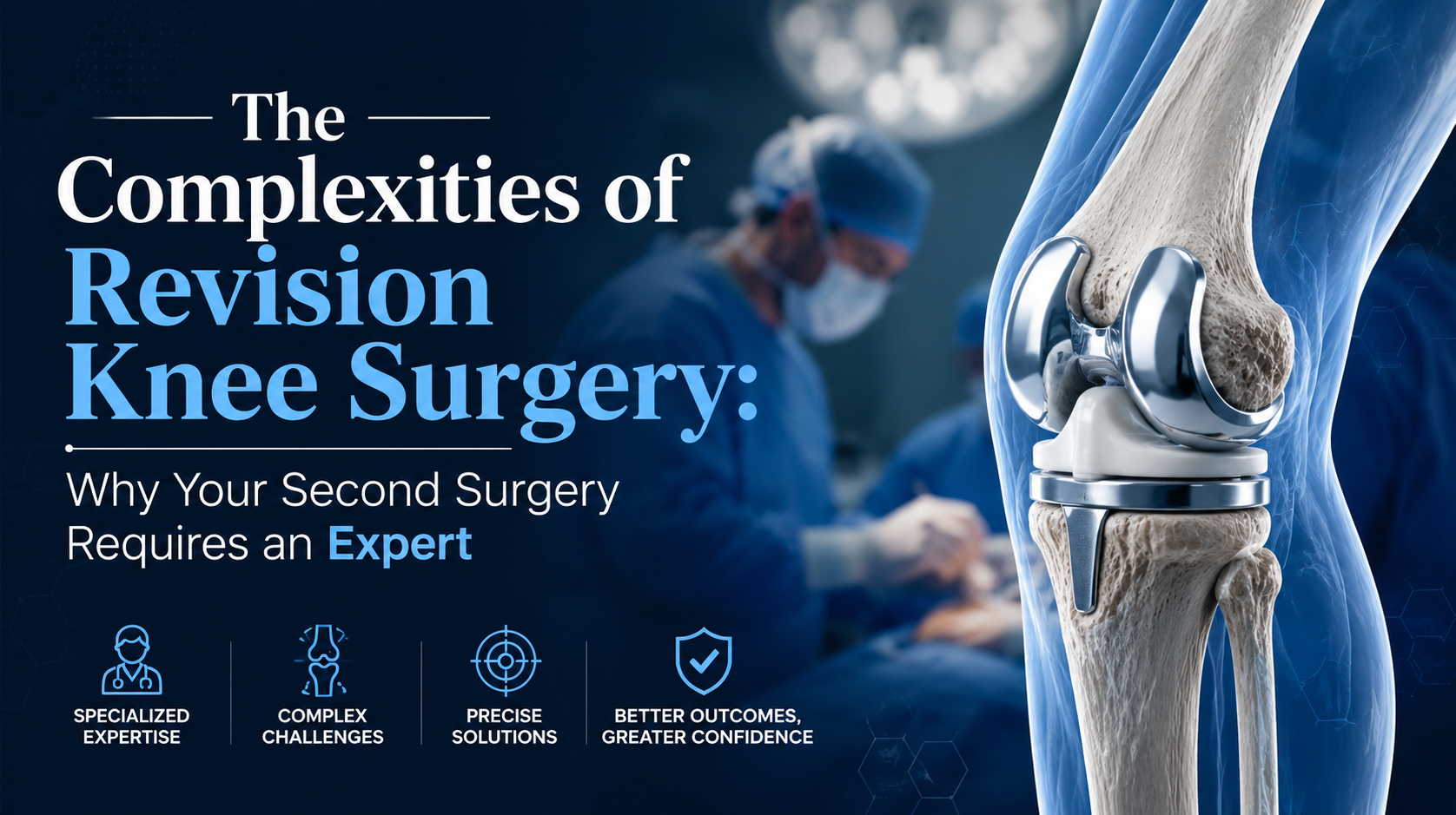
Implant malalignment, abnormal patellar tracking, or excessive soft-tissue tightness can result in arthrofibrosis—an overproduction of dense scar tissue inside the joint capsule. Patients present with severe stiffness, persistent aching, and an inability to achieve full extension or adequate flexion despite rigorous physical therapy. The Complexities of Revision Knee Surgery: Why Your Second Surgery Requires an Expert
Evaluating a failing primary knee replacement requires a structured, multi-step diagnostic approach. The objective is to differentiate between septic and aseptic loosening, evaluate remaining bone stock, and plan structural restoration.
[ CLINICAL EVALUATION ] ---> [ SEROLOGICAL SCREENING ] ---> [ JOINT ASPIRATION ] ---> [ ADVANCED 3D IMAGING ]
Physical exam, stance, ESR, CRP, & D-Dimer Synovial WBC count, CT with Metal Artifact
gait, & scar inspection to screen for infection % PMN, & alpha-defensin Reduction (MARS) & X-rays
Every patient undergoing evaluation for a secondary knee operation undergoes blood testing, including Erythrocyte Sedimentation Rate (ESR) and C-Reactive Protein (CRP). If inflammatory markers are elevated, a joint aspiration is performed under sterile conditions. Synovial fluid is analyzed for:
Total white blood cell (WBC) count and differential (percentage of polymorphonuclear neutrophils).
Leukocyte esterase strip testing.
Alpha-defensin biomarker immunoassay, which provides high sensitivity and specificity for periprosthetic infection.
Aerobic, anaerobic, and fungal cultures to guide targeted antibiotic selection.
Standard long-leg standing X-rays assess overall mechanical alignment, limb length discrepancies, and joint line position. However, conventional radiographs often underestimate bone loss obscured by metal implants. Advanced Computed Tomography (CT) with Metal Artifact Reduction Software (MARS) allows the revision surgeon to assess osteolytic defects in 3D, measure rotational component alignment, and select appropriate bone reconstructive hardware prior to entering the operating room.
When revision total knee replacement surgery is required due to infection, the surgical strategy depends on the duration of symptoms, the infecting microorganism, and soft-tissue integrity.
+----------------------------------------------------------------------------------------------------+
| SINGLE-STAGE VS. TWO-STAGE PROTOCOL |
+----------------------------------+----------------------------------+------------------------------+
| Clinical Factor | Single-Stage Revision Protocol | Two-Stage Revision Protocol |
+----------------------------------+----------------------------------+------------------------------+
| Indication Criteria | Known, low-virulence organism | High-virulence, resistant |
| | with healthy soft-tissue envelope| organisms or unknown pathogens|
+----------------------------------+----------------------------------+------------------------------+
| Surgical Interventions | Explantation, debridement, and | Stage 1: Explantation & spacer|
| | direct reimplantation in one setting| Stage 2: Re-implantation later|
+----------------------------------+----------------------------------+------------------------------+
| Antibiotic Delivery | Systemic antibiotics post-op | High-dose antibiotic cement |
| | based on sensitivity culture | spacer plus systemic therapy |
+----------------------------------+----------------------------------+------------------------------+
| Interval Between Surgeries | None (Single surgical event) | 6 to 12 weeks of therapy |
| | | between Stage 1 and Stage 2 |
+----------------------------------+----------------------------------+------------------------------+
In high-virulence or chronic periprosthetic joint infections, two-stage revision remains the gold standard treatment:
Stage 1 (Explantation & Debridement): All primary components, bone cement, and infected soft tissues are removed. A custom or pre-formed articulating antibiotic-impregnated bone cement spacer is placed. This spacer releases local antibiotics (such as vancomycin and tobramycin) directly into the joint space while maintaining soft-tissue tension and joint mobility.
Interim Therapy: The patient receives 6 to 8 weeks of targeted intravenous or oral antibiotic therapy overseen by infectious disease specialists, monitored via weekly ESR and CRP levels.
Stage 2 (Reimplantation): Once clinical signs and inflammatory markers normalize, a second joint aspiration confirms clearance of the infection. The antibiotic spacer is then removed, and a definitive revision knee prosthesis is implanted. The Complexities of Revision Knee Surgery: Why Your Second Surgery Requires an Expert
One of the most complex aspects of revision knee surgery is managing bone loss caused by osteolysis, stress shielding, or component removal. The Anderson Orthopaedic Research Institute (AORI) classification system guides reconstructive decision-making:
[ AORI BONE DEFECT CLASSIFICATION ]
|
+---------------------------------------+---------------------------------------+
| | |
[ Type I: Intact Metaphysis ] [ Type II: Damaged Metaphysis ] [ Type III: Deficient Metaphysis ]
| | |
- Minor bone loss - Loss of cancellous bone - Complete metaphyseal collapse
- Standard cement/corticocancellous - Requires structural augments, - Demands zone 2/3 fixation via
grafting sufficient titanium cones, or sleeves highly constrained / custom implants
+----------------------------------------------------------------------------------------------------+
| AORI BONE LOSS RECONSTRUCTION |
+-----------------------+------------------------------------+---------------------------------------+
| AORI Classification | Defect Characteristics | Reconstructive Technique |
+-----------------------+------------------------------------+---------------------------------------+
| Type I | Minor, contained bone loss with | Bone cement fill, particulate bone |
| | preserved metaphyseal bone stock | graft, or autograft morselization |
+-----------------------+------------------------------------+---------------------------------------+
| Type IIA / IIB | Loss of metaphyseal cancellous | Metal block augments, porous titanium |
| | bone (unicondylar or bicondylar) | metaphyseal cones, or offset sleeves |
+-----------------------+------------------------------------+---------------------------------------+
| Type III | Severe bone loss involving an | Highly porous tantalum cones, custom |
| | entire condyle or metaphyseal segment| structural allografts, or distal femoral|
| | | replacement (hinged prostheses) |
+-----------------------+------------------------------------+---------------------------------------+
Historically, large bone defects were managed using structural allografts or thick bone cement blocks, which carried risks of non-union, wear, or early loosening. Modern revision knee surgery utilizes 3D porous titanium metaphyseal cones and sleeves.
+-----------------------+
| Joint Line Level | <--- Zone 1: Epiphyseal Fixation
+-----------------------+ (Restored with modular augments)
| Metaphyseal Sleeve | <--- Zone 2: Metaphyseal Fixation
| or Titanium Cone | (Primary load-bearing zone)
+-----------------------+
| Intramedullary Stem | <--- Zone 3: Diaphyseal Fixation
| (Fluted / Offset) | (Provides axial stability)
+-----------------------+
These highly porous structures mimic native trabecular bone geometry, promoting rapid bone ingrowth. By securing fixation in the metaphyseal zone (Zone 2), the implant transfers weight-bearing forces directly to healthy bone, shielding weaker epiphyseal areas and providing long-term structural stability.
Re-establishing accurate knee kinematics during revision total knee replacement surgery requires careful reconstruction of joint alignment, joint line position, and stability.
[ ELEVATED JOINT LINE HAZARD ] ===> Pseudopatella Baja ===> Anterior Knee Pain & Range-of-Motion Deficit
[ RESTORED JOINT LINE HEIGHT ] ===> Balanced Collaterals ===> Smooth Flexion/Extension Mechanics
During primary implant explantation, bone loss can lead to an unintended elevation of the joint line if the surgeon simply uses thicker polyethylene inserts to fill the space. Elevating the joint line creates pseudopatella baja, leading to anterior knee pain, restricted flexion, patellar impingement, and mid-flexion instability.
To restore the true anatomic joint line, the revision surgeon uses distal femoral augments to lower the femoral component, ensuring proper patellofemoral tracking and balanced collateral ligament tension throughout the arc of motion. The Complexities of Revision Knee Surgery: Why Your Second Surgery Requires an Expert
Because collateral ligaments are often stretched, compromised, or absent in revision cases, selecting the correct level of mechanical constraint is vital:
+----------------------------------------------------------------------------------------------------+
| PROSTHETIC CONSTRAINT HIERARCHY |
+----------------------------------+-----------------------------------------------------------------+
| Constraint Level | Indications & Biomechanical Characteristics |
+----------------------------------+-----------------------------------------------------------------+
| Posterior-Stabilized (PS) | Intact collateral ligaments with competent soft-tissue envelope;|
| | used in minor, uncomplicated revision procedures. |
+----------------------------------+-----------------------------------------------------------------+
| Non-Spigoted Condylar Constrained| Mild to moderate collateral ligament laxity or asymmetrical gap |
| Knee (CCK) | imbalances; wide spine-and-cam mechanism controls stability. |
+----------------------------------+-----------------------------------------------------------------+
| Rotating Hinge Knee (RHK) | Severe ligament loss, global instability, AORI Type III bone |
| | defects, or complete medial collateral ligament (MCL) disruption|
+----------------------------------+-----------------------------------------------------------------+
Using a Rotating Hinge Knee (RHK) provides mechanical stability while allowing controlled axial rotation, reducing torsional stress on the bone-implant interface and protecting surrounding bone stock.
Modern technology has significantly improved the predictability and accuracy of complex revision joint replacement.
[ PREOP 3D CT MODELING ] ---> [ ROBOTIC / NAVIGATED MAPPING ] ---> [ PRECISION BONE RESECTION ] ---> [ CUSTOM IMPLANT FIT ]
Calculates bone loss, Tracks anatomical landmarks Executes sub-millimeter cuts Secures zonal fixation
joint line, & rotational and dynamic soft-tissue tension around remaining bone stock with minimal bone removal
alignment requirements in real time
Incorporating robotic-assisted total knee arthroplasty protocols into revision procedures allows surgeons to map structural deformities in real time. Optical tracking arrays collect spatial data on remaining bone stock, joint line height, and collateral ligament laxity. The robotic system then provides real-time guidance during bone preparation, helping preserve healthy bone and align new components accurately.
In cases involving severe structural bone loss or prior traumatic deformities, off-the-shelf revision implants may not provide adequate stability. Advanced 3D printing technology enables the fabrication of patient-specific custom titanium implants. Designed from preoperative CT data, these custom constructs fit exact defect boundaries, restoring joint function in complex reconstructive cases.
Rehabilitation following revision knee surgery requires a patient-specific, multi-phase physical therapy program tailored to the extent of bone reconstruction and ligament stability.
+----------------------------------------------------------------------------------------------------+
| REVISION KNEE REHABILITATION TIMELINE |
+-----------------------+------------------------------------+---------------------------------------+
| Recovery Phase | Therapeutic Focus & Interventions | Functional Milestones |
+-----------------------+------------------------------------+---------------------------------------+
| Phase 1: Inpatient | Multimodal pain management, passive| Safe bed mobility, toe-touch to partial|
| (Days 0 to 4) | motion, and edema control | weight-bearing with walker support |
+-----------------------+------------------------------------+---------------------------------------+
| Phase 2: Early Rehab | Active quadriceps activation and | Controlled knee extension (0°) and |
| (Weeks 2 to 6) | progressive gait training | flexion reaching 90° ROM |
+-----------------------+------------------------------------+---------------------------------------+
| Phase 3: Advanced | Balance retraining, stairs, and | Transition to unassisted walking and |
| (Weeks 7 to 16) | functional muscular endurance | return to daily independent activities|
+-----------------------+------------------------------------+---------------------------------------+
Protected Weight-Bearing: When extensive bone grafting, metaphyseal cones, or hinged implants are used, patients may follow a protected weight-bearing protocol (using a walker or crutches) for 4 to 6 weeks to allow full osseointegration of porous metal components.
Multimodal Analgesia and Thromboprophylaxis: Pain management utilizes targeted regional nerve blocks (such as adductor canal blocks) alongside multimodal oral medications, minimizing opioid reliance. Mechanical compression devices and tailored pharmacological anticoagulation protect against deep vein thrombosis (DVT).
Targeted Gait Retraining: Dedicated physical therapy helps correct compensatory walking habits formed during years of joint dysfunction, building quadriceps strength and improving long-term stability.
A revision total knee replacement surgery is significantly more complex and typically takes longer than a primary procedure. While a first-time knee replacement usually takes 1 to 2 hours, a revision surgery can take 2 to 4 hours (or more) depending on the amount of bone loss, scar tissue, and the difficulty of removing the old implants. The Complexities of Revision Knee Surgery: Why Your Second Surgery Requires an Expert
Pain levels can vary from patient to patient. While the surgical site undergoes more extensive work during a revision total knee replacement surgery, modern multimodal pain management techniques used by an expert orthopedics surgeon specialist in Gurgaon ensure that post-operative discomfort is carefully controlled and managed. The Complexities of Revision Knee Surgery: Why Your Second Surgery Requires an Expert
When performed by a highly skilled specialist, the success rate of a secondary knee operation is remarkably high. However, because it is a complex intervention, choosing the best orthopedics surgeon specialist in Gurgaon like Dr. Hemant Sharma drastically reduces the risk of complications and ensures a much more predictable, long-lasting outcome. The Complexities of Revision Knee Surgery: Why Your Second Surgery Requires an Expert
Modern revision implants are incredibly durable. While they may not always last quite as long as a primary implant due to underlying bone changes, a properly executed revision total knee replacement surgery can comfortably last 10 to 15 years or longer, depending on the patient’s lifestyle, activity levels, and overall bone health. The Complexities of Revision Knee Surgery: Why Your Second Surgery Requires an Expert
The most common warning signs include persistent or worsening pain, a feeling of instability (the knee “giving way”), swelling, stiffness, or a noticeable change in the alignment of the leg. If you experience these symptoms, you should consult an orthopedics surgeon specialist in Gurgaon immediately for a diagnostic evaluation. The Complexities of Revision Knee Surgery: Why Your Second Surgery Requires an Expert
#Knee Revision Surgery #Revision Total Knee Replacement Surgery #Dr Hemant Sharma #Orthopedic Surgeon Gurgaon #Knee Replacement India #Joint Replacement Expert #Gurgaon Healthcare #Orthopedics Specialist #Knee Pain Relief #Mobility Restored