Dr. Hemant Sharma
FRCS (England) MRCS (England) DNB
Specialist in Revision Lower Limb ArthroPlasty
Complex Hip, Knee and Sports Injury

When you roll your ankle, you expect a few weeks of rest, some ice, and a gradual return to your daily routine. However, if your doctor diagnoses you with a syndesmotic injury, you are facing a completely different situation. Understanding the biomechanics of this injury reveals exactly why bone separation makes high ankle sprains a recovery nightmare.
Unlike a standard inversion injury that stretches the ligaments on the outside of your foot, a high sprain compromises the critical structural network binding your lower leg bones together. When these bones separate, even by a few millimeters, the entire stability of your lower body is compromised, turning a common injury into a prolonged, frustrating clinical ordeal.
To appreciate why bone separation makes high ankle sprains a recovery nightmare, it is essential to understand the unique anatomy of the lower leg. Your shin contains two parallel bones: the larger, weight-bearing tibia and the smaller, outer fibula.
These bones are bound together by the syndesmosis—a high-tensile network of ligaments and an interosseous membrane that acts as a structural shock absorber. A high ankle sprain occurs when a sudden, external rotation forces the foot upward and outward, tearing this membrane and forcing the tibia and fibula to physically widen. This specific bone separation disrupts the ankle mortise, meaning the socket no longer holds the ankle bone securely, completely destabilizing your structural foundation.
The primary reason why bone separation makes high ankle sprains a recovery nightmare boils down to simple physics and mechanical weight-bearing stress. When you suffer a low ankle sprain, the lateral ligaments are resting while you walk straight forward. However, with a high ankle sprain, the mechanics change completely:
The Spreading Effect: Every single time your heel hits the ground, your body weight pushes down, acting like a wedge that forces the tibia and fibula to spread outward.
Micro-Tearing Cycle: This continuous outward pressure pulls apart the newly forming collagen strands attempting to knit the syndesmosis back together.
Compounded Healing Times: Because everyday mobility constantly re-tears the healing tissue, the recovery timeline is instantly doubled or tripled compared to standard lateral sprains.
This relentless mechanical widening explains why bone separation makes high ankle sprains a recovery nightmare for active individuals. Pushing through the pain doesn’t build resilience; it simply prevents the skeletal gap from closing. To know more about Why Bone Separation Makes High Ankle Sprains a Recovery Nightmare. consult Dr Hemant Sharma Today.
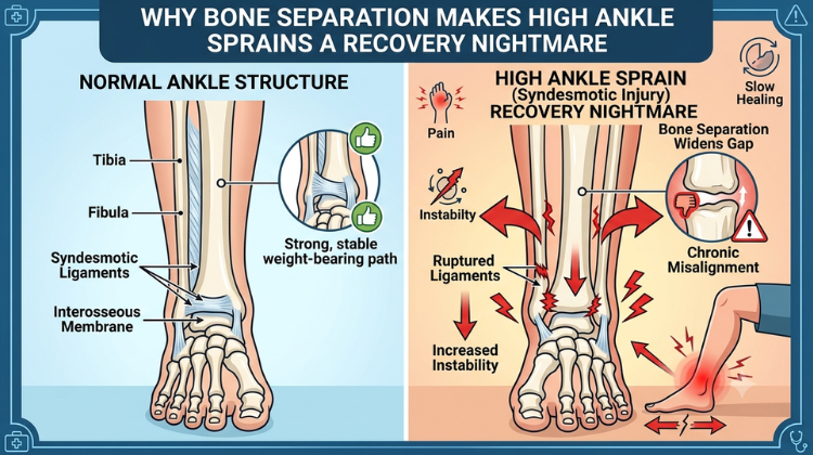
Ignoring the widening between your lower leg bones leads to permanent structural issues. If the tibia and fibula do not heal in tight, parallel alignment, the ankle joint will experience abnormal friction, shifting your entire biomechanical alignment. Over time, this chronic instability leads to early-onset joint degeneration, persistent micro-swelling, and a permanent reduction in your athletic agility. This long-term vulnerability is another reason why bone separation makes high ankle sprains a recovery nightmare when proper immobilization protocols are skipped early on.
Also Read: High Ankle Sprain vs. Normal Sprain: Why the Syndesmosis Takes Twice as Long to Heal
Managing a syndesmotic widening and preventing long-term joint damage requires an exceptionally precise clinical diagnosis. When dealing with complex ligament separations, seeking an expert medical evaluation is the safest way to ensure a complete, stable recovery.
Current Clinical Role: Chairman of Orthopaedics and Joint Replacement at Marengo Asia Hospitals, Gurugram.
Decades of Expertise: Over 31 years of global clinical practice, including 11 years of advanced surgical training and practice within the UK National Health Service (NHS).
Elite Professional Credentials: Holds an MBBS alongside prestigious certifications as a Fellow of the Royal College of Surgeons (FRCS, Trauma & Orthopaedics, UK) and Member of the Royal College of Surgeons (MRCS, England).
Robotic Surgery Pioneer: Recognized as a premier first-generation robotic-assisted orthopedic surgeon in India, having successfully completed more than 500 advanced robotic procedures.
Award-Winning Excellence: Awarded the highly distinguished Service Excellence Award (2025) for Robotic Orthopaedics Surgery & Joint Replacement at the Times Network India Health Summit.
Getting an accurate assessment from an internationally trained authority like Dr. Hemant Sharma, the best ortho surgeon in Gurgaon, guarantees that any structural bone separation is accurately mapped, managed, and treated using world-class protocols.
Ultimately, overcoming this injury requires patience and strict adherence to medical advice. Because bone separation makes high ankle sprains a recovery nightmare, treating it like a standard twist will only result in chronic pain. Restoring structural integrity requires early non-weight-bearing protocols, rigid immobilization using a walking boot, and progressive physical therapy to strengthen the surrounding calf muscles.
By respecting the unique biomechanics of the syndesmosis and seeking guidance from top-tier specialists like Dr. Hemant Sharma, the best ortho surgeon in Gurgaon, you can ensure your leg bones heal perfectly aligned—effectively ending the recovery nightmare and restoring your peak performance.
High Ankle Sprain Recovery: Biomechanics, Clinical Evaluation, Advanced Surgical Interventions, and Rehabilitation Protocols
A high ankle sprain—clinically recognized as a syndesmotic ankle injury—is widely regarded as one of the most challenging musculoskeletal conditions in orthopedic medicine. Unlike standard lateral ankle twists that involve extra-articular ligaments below the joint line, a high sprain compromises the syndesmotic complex connecting the tibia (shinbone) and fibula (outer leg bone).
[ HIGH ANKLE TRAUMA MECHANISM & PATHWAY ]
|
+-----------------------------------------+-----------------------------------------+
| |
[ EXTERNAL ROTATION & FORCED DORSIFLEXION ] [ MECHANICAL MORTISE DISRUPTION ]
| |
- Outward Foot Rotation Relative to Lower Leg - Distal Tibiofibular Widening (> 2 mm)
- Ruptures AITFL, PITFL, & Interosseous Membrane - Talus Shifts Laterally Under Loading
- Expands Ankle Mortise Socket Dimensions - Concentrates Contact Stress on Cartilage
|
v
[ CLINICAL MANAGEMENT PATHWAY ]
- Diagnostic Stress Testing (Squeeze & External Rotation)
- Weight-Bearing Radiographs & High-Resolution MRI
- Non-Surgical Rigid Immobilization vs. Dynamic Fixation
Because this ligamentous network forms the socket holding the talus (the ankle mortise), damage to these tissues alters lower-leg kinematics. Weight-bearing forces place direct strain on healing syndesmotic tissues, making the recovery process significantly longer than that of a standard inversion sprain. Without precise evaluation and specialized care, an unhealed syndesmotic tear can lead to persistent joint instability, altered foot mechanics, and accelerated post-traumatic osteoarthritis. To know more about Why Bone Separation Makes High Ankle Sprains a Recovery Nightmare. consult Dr Hemant Sharma Today.
Understanding why a high sprain demands an extended recovery period requires a comparison of the distinct ligament systems involved in high ankle sprain vs. normal sprain mechanisms.
+-------------------------------------------------------------------------------------------------------------------+
| LIGAMENT ANATOMY & BIOMECHANICAL MATRIX |
+----------------------------------+---------------------------------------+----------------------------------------+
| Structural Dimension | Low Ankle Sprain (Lateral Complex) | High Ankle Sprain (Syndesmotic Complex)|
+----------------------------------+---------------------------------------+----------------------------------------+
| Primary Ligaments Involved | ATFL (Anterior Talofibular) & | AITFL, PITFL, Transverse Ligament, and |
| | CFL (Calcaneofibular Ligament) | Interosseous Membrane (IOM) |
+----------------------------------+---------------------------------------+----------------------------------------+
| Primary Injury Mechanism | Plantarflexion with inward foot roll | External rotation of foot relative to |
| | (inversion strain on lateral wall) | leg with forced dorsiflexion |
+----------------------------------+---------------------------------------+----------------------------------------+
| Primary Structural Role | Limits excessive inversion and | Binds tibia and fibula together; |
| | anterior translation of the talus | maintains structural ankle mortise |
+----------------------------------+---------------------------------------+----------------------------------------+
| Weight-Bearing Strain Dynamic | Low direct axial widening force; | High axial widening force; weight |
| | motion occurs primarily along planes | forces tibia and fibula to spread apart|
+----------------------------------+---------------------------------------+----------------------------------------+
The standard lateral complex consists of three key ligaments that stabilize the outer ankle joint:
Anterior Talofibular Ligament (ATFL): The primary restrictor against anterior talar translation and internal rotation. Because it is the weakest lateral structure, it is the first tissue disrupted during an inward foot twist.
Calcaneofibular Ligament (CFL): Passes across the subtalar joint, stabilizing the calcaneus against excessive inversion tilt.
Posterior Talofibular Ligament (PTFL): A heavy posterior band that resists backward displacement of the talus, rarely damaged in simple twists.
These ligaments lie outside the main weight-bearing column of the leg. As a result, walking exerts minimal direct separation force on repairing lateral fibers once acute inflammation subsides.
[ SYNDESMOTIC "ZIPPER" ARCHITECTURE ]
Tibia (Shinbone) Fibula (Outer Bone)
+------------------+ +--------------------+
| |====================================| |
| | Interosseous Membrane (IOM) | |
| |====================================| |
| | AITFL (Anterior Inferior) | |
| |------------------------------------| |
+------------------+ +--------------------+
\ /
\--- Ankle Mortise Socket (Talus) -/
The syndesmosis acts as a high-tensile fibrous joint connecting the distal tibia and fibula. This complex includes:
Anterior Inferior Tibiofibular Ligament (AITFL): Spans the front aspect of the distal tibiofibular joint.
Posterior Inferior Tibiofibular Ligament (PITFL) & Transverse Ligament: Heavy posterior structures providing deep structural support to the ankle socket.
Interosseous Membrane (IOM) & Interosseous Ligament (IOL): Broad sheets of high-tensile fibrous tissue extending up the lower leg between the tibia and fibula.
Together, these structures form a secure socket—the ankle mortise—that holds the talus bone in place. When the syndesmosis is torn, the tibia and fibula separate under weight-bearing loads, disturbing normal joint alignment. To know more about Why Bone Separation Makes High Ankle Sprains a Recovery Nightmare. consult Dr Hemant Sharma Today.
The anatomical distinction explains why recovery times vary significantly between high and low ankle sprains.
[ AXIAL WEIGHT DISTRIBUTION MECHANICS ]
|
+-------------------------------------------+-------------------------------------------+
| |
[ STEPPING ON A LOW SPRAIN ] [ STEPPING ON A HIGH SPRAIN ]
| |
- Load travels axially through ankle joint - Talus acts as a rigid wedge within mortise
- Lateral ligaments stay relatively relaxed - Forces tibia and fibula to spread apart
- Minimally disrupts healing ATFL collagen fibers - Repeatedly stretches torn syndesmotic tissue
|
v
[ RECOVERY TIMELINE IMPLICATION ]
- Low Sprain: Accelerated functional rehab (2–6 weeks)
- High Sprain: Prolonged strict protection (8–16+ weeks)
The upper surface of the talus bone is wider at the front than at the back. When the foot flexes upward during walking (dorsiflexion), this wider front section moves back into the ankle mortise. If the syndesmotic ligaments are torn, the talus acts as a wedge, forcing the tibia and fibula apart with every step. This separation disrupts healing tissue and delays structural recovery.
Lateral ankle ligaments benefit from a rich surrounding vascular network that promotes localized blood flow and cellular migration. In contrast, the interosseous membrane and syndesmotic ligaments have a sparse blood supply. This limited blood flow slows collagen synthesis, extending the timeline required for the syndesmosis to regain its structural integrity. To know more about Why Bone Separation Makes High Ankle Sprains a Recovery Nightmare. consult Dr Hemant Sharma Today.
Distinguishing a high ankle sprain from a low ankle sprain requires systematic physical testing and appropriate imaging techniques.
+----------------------------------------------------------------------------------------------------+
| CLINICAL DIAGNOSTIC EVALUATION MATRIX |
+-----------------------+------------------------------------+---------------------------------------+
| Diagnostic Test | Procedure Method | Positive Indicator for High Sprain |
+-----------------------+------------------------------------+---------------------------------------+
| Squeeze Test | Squeezing mid-calf (tibia and | Pain radiating down to the distal |
| | fibula) at mid-leg height | syndesmosis above the ankle joint |
+-----------------------+------------------------------------+---------------------------------------+
| External Rotation | Stabilizing lower leg while | Sharp pain felt above the ankle joint |
| Stress Test | rotating foot outward | as the talus spreads the mortise |
+-----------------------+------------------------------------+---------------------------------------+
| Cotton Test | Translating talus laterally inside | Excessive lateral movement or clunking|
| (Intraoperative) | the ankle mortise | indicating mortise instability |
+-----------------------+------------------------------------+---------------------------------------+
| Weight-Bearing | Bilateral standing anteroposterior | Medial clear space > 4 mm or |
| Radiographs | (AP) and mortise views | tibiofibular clear space > 6 mm |
+-----------------------+------------------------------------+---------------------------------------+
[ DIAGNOSTIC EVALUATION PATHWAY ]
|
+---> Physical Tests: Squeeze Test & External Rotation Stress Test
|
+---> Standing Stress Radiographs (Assess Medial / Tibiofibular Clear Space)
|
+---> High-Resolution Contrast MRI (Evaluate AITFL, PITFL, and IOM Integrity)
The Squeeze Test: Compressing the mid-calf compresses the upper tibia and fibula, causing the lower ends of the bones to bow outward. Pain felt at the distal lower leg during this test indicates syndesmotic damage.
External Rotation Stress Test: With the knee flexed at 90 degrees, the foot is gently rotated outward relative to the leg. Recreating pain in the anterior lower leg indicates a high sprain.
Standard non-weight-bearing X-rays can miss mild-to-moderate syndesmotic injuries. Accurate evaluation requires:
Weight-Bearing Stress Radiographs: Standing X-rays highlight subtle joint widening under load. A tibiofibular clear space greater than 6 mm or a medial clear space greater than 4 mm suggests syndesmotic instability.
Magnetic Resonance Imaging (MRI): The standard imaging method for evaluating soft tissues. High-resolution MRI visualizes torn AITFL, PITFL, and interosseous membrane fibers, helping clinicians grade the injury and plan appropriate treatment. To know more about Why Bone Separation Makes High Ankle Sprains a Recovery Nightmare. consult Dr Hemant Sharma Today.
Treatment choices depend on the structural severity of the ligament tear and the presence of joint instability.
+----------------------------------------------------------------------------------------------------+
| SYNDESMOTIC INJURY GRADING |
+-------------------+------------------------------------+-------------------------------------------+
| Severity Grade | Anatomical & Structural Condition | Recommended Treatment Approach |
+-------------------+------------------------------------+-------------------------------------------+
| Grade I | Microscopic AITFL tearing; no | Rigid cast/boot immobilization for 4 to 6 |
| (Mild) | widening or structural instability | weeks, followed by physical therapy |
+-------------------+------------------------------------+-------------------------------------------+
| Grade II | Partial AITFL & IOM tearing; | Protected weight-bearing or dynamic |
| (Moderate) | latent instability under stress | surgical stabilization if unstable |
+-------------------+------------------------------------+-------------------------------------------+
| Grade III | Complete disruption of AITFL, | Surgical stabilization (TightRope suture |
| (Severe) | PITFL, and IOM; frank dislocation | button or syndesmotic screws) required |
+-------------------+------------------------------------+-------------------------------------------+
[ GRADE I (STABLE) ] ===> Non-Weight-Bearing Immobilization -> Progressive Rehabilitation
[ GRADE II (LATENT) ] ===> Stress Radiograph / Arthroscopy -> Targeted Stabilization
[ GRADE III (UNSTABLE) ] ===> Surgical Reduction & Fixation (Suture-Button or Transfixation Screws)
Stable Grade I syndesmotic injuries require protection from rotational forces. Unlike low ankle sprains—which benefit from early functional movement—a high ankle sprain typically requires an initial period of non-weight-bearing cast or rigid boot immobilization for 4 to 6 weeks to allow the torn fibers to heal without stretching. To know more about Why Bone Separation Makes High Ankle Sprains a Recovery Nightmare. consult Dr Hemant Sharma Today.
When complete ligament tearing allows the tibia and fibula to separate, surgical stabilization is necessary to restore joint alignment and preserve long-term ankle function.
When surgical stabilization is necessary, orthopedic surgeons choose between traditional rigid screws and modern flexible fixation devices.
+----------------------------------------------------------------------------------------------------+
| SURGICAL FIXATION COMPARISON MATRIX |
+----------------------------------+--------------------------------+--------------------------------+
| Surgical Parameter | Rigid Metallic Transfixation | Flexible Dynamic Suture-Button |
| | Screws (3.5 mm / 4.5 mm) | (e.g., Arthrex TightRope) |
+----------------------------------+--------------------------------+--------------------------------+
| Mechanical Rigidity | Complete rigid fixation; | Semi-rigid dynamic fixation; |
| | eliminates all micro-motion | permits natural fibular movement|
+----------------------------------+--------------------------------+--------------------------------+
| Second Surgery Requirement | Usually requires removal at 8– | Permanent implant; routine |
| | 12 weeks before full loading | hardware removal not required |
+----------------------------------+--------------------------------+--------------------------------+
| Physiological Joint Alignment | Risk of over-tightening the | Allows natural alignment of the|
| | syndesmosis during insertion | fibula within the tibial groove|
+----------------------------------+--------------------------------+--------------------------------+
| Postoperative Weight-Bearing | Delayed until screw removal to | Earlier weight-bearing in a |
| Timeline | prevent implant breakage | protective walking boot |
+----------------------------------+--------------------------------+--------------------------------+
[ RIGID TRANSMALLEOLAR SCREW ] [ DYNAMIC SUTURE-BUTTON SYSTEM ]
+------------------------------+ +----------------------------------+
| Transfixes Tibia & Fibula | | High-Strength FiberWire Suture |
| Rigidly; Risk of Metal Fatigue| vs | Tightened Between Metallic Buttons|
| Requires Second Removal Step | | Preserves Micro-Motion Dynamics |
+------------------------------+ +----------------------------------+
Transfixation screws pass through three or four bone cortices to hold the tibia and fibula together. While effective at stabilizing the joint, rigid screws restrict the natural rotation of the fibula during walking. As a result, patients often require a second procedure at 8 to 12 weeks to remove the hardware before resuming full activity and sports.
Modern surgical approaches frequently utilize flexible suture-button systems (such as the Arthrex TightRope). This implant uses a strong FiberWire suture suspended between two titanium buttons placed against the outer bone walls.
Dynamic Motion: Maintains accurate reduction of the ankle mortise while allowing natural fibular movement during gait.
No Routine Hardware Removal: Eliminates the need for a second surgery to remove metalwork, allowing for earlier weight-bearing and faster functional rehabilitation.
Failing to recognize or properly treat a high ankle sprain can lead to long-term joint complications.
[ PATHWAY TO POST-TRAUMATIC ARTHROSIS ]
|
+---------------------------------------+---------------------------------------+
| |
[ CHRONIC MORTISE WIDENING ] [ ALTERED CARTILAGE LOADING ]
| |
- Unhealed Syndesmosis Leaves Lateral Gap - Reduced Talofibular Contact Area
- Talus Shifts Lateral by Just 1 mm - Joint Contact Pressures Spike by 42%
- Causes Uneven Weight Distribution Across Cartilage - Accelerates Chondrocyte Loss & Erosion
|
v
[ POST-TRAUMATIC OSTEOARTHRITIS ]
- Chronic Ankle Pain & Recurrent Swelling
- Progressive Stiffness & Joint Space Narrowing
- May Require Ankle Arthrodosis or Arthroplasty
Studies show that a 1-millimeter lateral shift of the talus reduces the contact area between the tibia and talus by up to 42%. This reduction concentrates weight-bearing forces onto a smaller surface area of cartilage, accelerating joint wear and increasing the risk of early-onset post-traumatic osteoarthritis.
Rehabilitation after a high ankle sprain requires a gradual, phased approach focused on restoring strength, joint position awareness (proprioception), and dynamic rotational stability.
Phase 1 (Weeks 0–4): [ Protected Phase ] ===> Non-weight-bearing in cast/boot; swelling control.
Phase 2 (Weeks 4–8): [ Mobility Phase ] ===> Gentle weight-bearing, sagittal-plane range of motion.
Phase 3 (Weeks 8–12): [ Strengthening Phase ] ===> Proprioceptive training, resistance bands, gait correction.
Phase 4 (Weeks 12+): [ Sports Return ] ===> Multi-directional agility drills, pivoting, sports clearance.
Phase I: Protection and Inflammation Control (Weeks 0–4) Focuses on protecting healing ligaments using a non-weight-bearing cast or rigid walking boot, along with elevation and gentle, non-weight-bearing range-of-motion exercises in the sagittal plane (flexing the foot up and down).
Phase II: Progressive Weight-Bearing and Mobility (Weeks 4–8) Gradually introduces weight-bearing as tolerated while protecting the ankle from outward rotational movements. Exercises focus on rebuilding calf strength and basic balance.
Phase III: Proprioceptive Retraining and Dynamic Stability (Weeks 8–12) Includes single-leg balance training, wobble board exercises, and functional movements to restore joint position sense and protect against re-injury.
Phase IV: Sport-Specific Agility and Pivoting (Weeks 12+) Gradually incorporates multi-directional agility drills, lateral cutting maneuvers, and sport-specific training once full strength and pain-free movement are achieved.
#High Ankle Sprain #Syndesmosis Injury #Ankle Bone Separation #Tibia And Fibula #Ankle Ligament Tear #Ankle Recovery Timeline #Ortho Surgeon Gurgaon #Dr Hemant Sharma #Marengo Asia Hospital #Best Orthopedic Doctor #Sports Injury Recovery #Ankle Rehabilitation #Ankle Joint Pain #Ligament Reconstruction #Trauma Surgeon India #FRCS Orthopaedics #Ankle Instability #Ankle Mortise Widening #Squeeze Test Ankle #External Rotation Injury #External Rotation Injury #Joint Preservation #Robotic Joint Surgery #Ankle Swelling #Calf Pain #Foot And Ankle Specialist #Ankle Brace Recovery #Sports Medicine India #Gurgaon Orthopaedic Clinic #Lower Leg Injury #Ankle Arthritis Prevention
A: Bone separation makes high ankle sprains a recovery nightmare because it compromises the syndesmotic ligaments holding the tibia and fibula together. Every time you stand or take a step, your body weight acts as a physical wedge that forces these two bones to spread apart. This continuous mechanical widening repeatedly pulls apart the newly healing tissue, making the recovery process exceptionally slow, painful, and prone to setbacks. To know more about Why Bone Separation Makes High Ankle Sprains a Recovery Nightmare. consult Dr Hemant Sharma Today.
A: If the gap between the tibia and fibula is left untreated, the ankle mortise remains widened and unstable. This structural instability alters your lower body biomechanics, causing the ankle joint to wear down unevenly. Over time, unmanaged bone separation leads to chronic joint pain, persistent swelling, localized weakness, and a highly elevated risk of early-onset ankle osteoarthritis. To know more about Why Bone Separation Makes High Ankle Sprains a Recovery Nightmare. consult Dr Hemant Sharma Today.
A: Dr. Hemant Sharma is widely recognized as the best ortho surgeon in Gurgaon for complex musculoskeletal trauma, advanced sports injuries, and joint reconstruction. Currently practicing as the Chairman of Orthopaedics at Marengo Asia Hospitals, Gurugram, he brings over 31 years of distinguished global clinical experience spanning leading medical centers across both India and the United Kingdom (NHS). To know more about Why Bone Separation Makes High Ankle Sprains a Recovery Nightmare. consult Dr Hemant Sharma Today.
A: Dr. Hemant Sharma holds elite international surgical credentials from the United Kingdom, including a fellowship as a Fellow of the Royal College of Surgeons (FRCS in Trauma & Orthopaedics, UK) and a membership certification (MRCS, England). He is also a celebrated pioneer of robotic-assisted orthopedic surgery in India, with a track record of over 500 successful robotic joint procedures and recipient of the 2025 Service Excellence Award. To know more about Why Bone Separation Makes High Ankle Sprains a Recovery Nightmare. consult Dr Hemant Sharma Today.
A: Mild cases without significant widening are managed with strict non-weight-bearing rest, rigid boot immobilization, and targeted physical therapy. However, if imaging reveals severe bone separation, surgical intervention is often required to stabilize the joint. Surgeons utilize specialized syndesmotic screws or a flexible “tightrope” fixation device to pull the tibia and fibula back into perfect alignment so the ligaments can heal securely. To know more about Why Bone Separation Makes High Ankle Sprains a Recovery Nightmare. consult Dr Hemant Sharma Today.