Opening: 09AM - 08PM

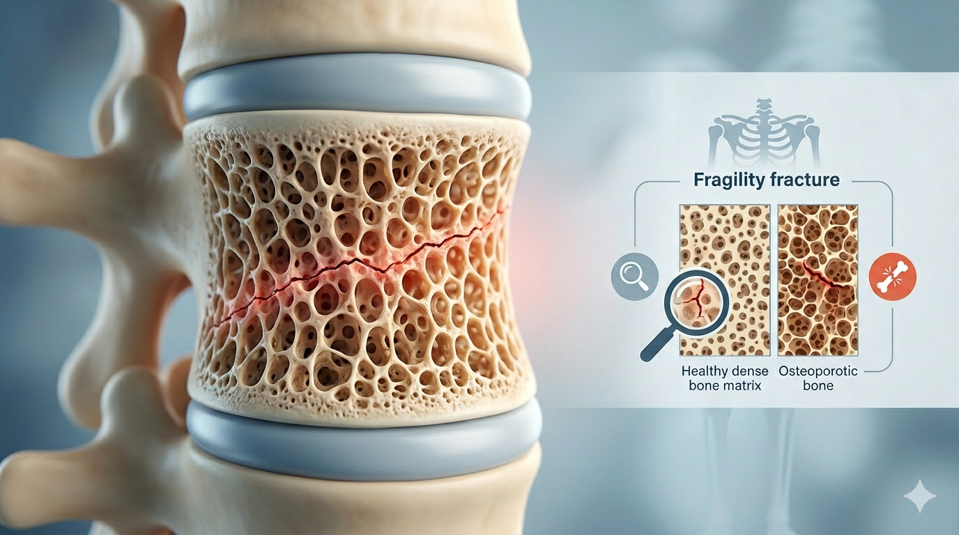
A fragility fracture is often the first warning sign that your bones are becoming weaker than they should be. Unlike fractures caused by major accidents or sports injuries, a fracture can occur after a minor fall, slip, or even routine daily activities. These fractures are commonly associated with osteoporosis and low bone density, making them a serious health concern, especially among older adults.

Understanding the causes, symptoms, and prevention strategies for a fracture is essential for maintaining long-term bone health and reducing the risk of future injuries. According to global health organizations, most fragility fractures occur due to weakened bones that cannot withstand normal stress or low-impact trauma.

What Is a Fragility Fracture?

TABLE OF CONTENT

A fragility fracture is a broken bone that occurs from low-energy trauma, such as a fall from standing height or less. Healthy bones generally do not break under such circumstances, but weakened bones affected by osteoporosis or other bone disorders are more vulnerable.

In many cases, a fracture serves as an early indicator of underlying bone weakness. Unfortunately, many individuals are unaware they have osteoporosis until they experience their first fragility fracture.

Why Does a Fragility Fracture Occur?

Several factors contribute to the development of a fragility fracture, including:

1. Osteoporosis

Osteoporosis reduces bone density and weakens bone structure, making fractures more likely. It is the leading cause of a fragility fracture.

2. Aging

As people age, bone mass naturally decreases, increasing the risk of fractures.

3. Hormonal Changes

Postmenopausal women face a higher risk due to declining estrogen levels, which affect bone strength.

4. Nutritional Deficiencies

Low calcium and vitamin D levels can weaken bones over time.

5. Sedentary Lifestyle

Lack of physical activity reduces bone strength and balance, increasing fracture risk.

Common Types of Fragility Fracture

A fragility fracture can occur in various parts of the body, but the most common locations include:

Hip Fractures

Hip fractures are among the most serious forms of fragility fracture and often require surgical intervention.

Spine (Vertebral) Fractures

These fractures may occur without a noticeable fall and can lead to chronic pain, height loss, and posture changes.

Wrist Fractures

A simple fall onto an outstretched hand can result in a wrist fragility fracture.

Shoulder Fractures

The upper arm bone near the shoulder is another common site for fracture in individuals with weakened bones.

Warning Signs of Weak Bones

Many people do not experience symptoms until a fracture occurs. However, warning signs may include:

  • Frequent back pain
  • Loss of height over time
  • Stooped posture
  • Fractures resulting from minor falls
  • Reduced mobility
  • General bone weakness

These symptoms may indicate osteoporosis and should prompt medical evaluation.


Who Is Most at Risk for a Fragility Fracture?

Certain individuals are more likely to experience a fracture, including:

  • Adults over 50 years of age
  • Postmenopausal women
  • Individuals with osteoporosis
  • People with a family history of fractures
  • Smokers
  • Heavy alcohol consumers
  • Individuals with low body weight
  • Patients taking long-term corticosteroid medications

Understanding these risk factors can help identify individuals who may benefit from early screening.

How Is a Fragility Fracture Diagnosed?

Doctors use several methods to diagnose a fracture and evaluate bone health:

Physical Examination

Assessment of pain, swelling, and mobility.

Imaging Tests

  • X-rays
  • CT scans
  • MRI scans

Bone Density Testing

A DEXA scan measures bone mineral density and helps diagnose osteoporosis. It is considered the gold standard for assessing fracture risk.

Treatment Options for Fragility Fracture

Treatment depends on the fracture location, severity, and overall health of the patient.

Non-Surgical Treatment

  • Casting or bracing
  • Pain management
  • Physical therapy
  • Calcium and vitamin D supplementation

Surgical Treatment

Some fractures, particularly hip and complex fractures, may require surgery to stabilize the bone and restore mobility.

Osteoporosis Management

Treating the underlying cause is crucial to preventing future fractures. This may include medications, lifestyle modifications, and nutritional support.

How to Prevent a Fragility Fracture

Preventing a fragility fracture starts with maintaining strong bones throughout life.

Eat a Bone-Healthy Diet

Consume adequate calcium, protein, and vitamin D.

Exercise Regularly

Weight-bearing and resistance exercises help strengthen bones and improve balance.

Avoid Smoking and Excessive Alcohol

Both habits negatively affect bone health.

Schedule Bone Density Screening

Early detection of osteoporosis can significantly reduce fracture risk.

Prevent Falls

Simple measures such as proper footwear, home safety modifications, and vision checks can lower the likelihood of falls.

Why Early Orthopedic Care Matters

Prompt evaluation and treatment after a fragility fracture can prevent complications and improve recovery outcomes. Individuals who suffer one fracture are at significantly higher risk of experiencing another fracture in the future.

For patients seeking expert orthopedic care in Gurgaon, consulting an experienced specialist is essential. Many patients trust Dr. Hemant best Orthopedics Surgeon Specialist in Gurgaon, recognized for advanced orthopedic treatments, fracture management, joint replacement procedures, and comprehensive bone health care.

Fragility fractures serve as a key clinical sign of underlying osteoporosis or underlying osteopenia. Without early diagnosis, surgical stabilization, and secondary prevention strategies, a single low-impact fracture often marks the start of a cascade of recurring fractures, progressive disability, and loss of independence.

Biomechanics and Pathophysiology of Bone Fragility

Bone strength depends on two main components: Bone Mineral Density (BMD) and Bone Quality.

+-------------------------------------------------------------------------------------------------------------------+
|                                     MECHANICAL & BIOLOGICAL BONE DYNAMICS                                         |
+----------------------------------+---------------------------------------+----------------------------------------+
| Bone Property Dimension          | Physiological Determinant             | Pathological Impact in Osteoporosis    |
+----------------------------------+---------------------------------------+----------------------------------------+
| Bone Mineral Density (BMD)       | Hydroxyapatite crystal mass per unit  | Progressive mineral loss leading to    |
|                                  | volume (measured via DEXA T-score)    | low bone density and osteopenia        |
+----------------------------------+---------------------------------------+----------------------------------------+
| Trabecular Microarchitecture     | Three-dimensional arrangement of       | Loss of trabecular connectivity and    |
|                                  | internal cancellous cross-struts      | conversion to disconnected rods        |
+----------------------------------+---------------------------------------+----------------------------------------+
| Cortical Geometry & Porosity     | Outer compact bone thickness and      | Endosteal resorption causing cortical  |
|                                  | Haversian canal pore density          | thinning and high intracortical voids  |
+----------------------------------+---------------------------------------+----------------------------------------+
| Organic Collagen Matrix          | Type I collagen cross-linking and     | Accumulation of Advanced Glycation     |
|                                  | micro-crack toughness                 | End-products (AGEs), reducing ductility|
+----------------------------------+---------------------------------------+----------------------------------------+

1. Uncoupled Bone Remodeling Dynamics

Throughout adult life, bone undergoes continuous remodeling through balanced osteoclast (resorption) and osteoblast (formation) activity. With advancing age, postmenopausal estrogen decline, or chronic systemic inflammation, this process becomes uncoupled:

  • Osteoclast activity increases, eroding deep pits within cancellous bone.

  • Osteoblast activity decreases, leaving microscopic voids unfilled.

Over time, this imbalance converts strong trabecular plates into thin, disconnected rods, drastically reducing the bone’s load-bearing capacity.

2. Cortical Thinning and Intracortical Porosity

While cancellous bone loss affects internal structure, cortical thinning directly reduces the bone’s resistance to bending and torsional forces. In postmenopausal women and elderly men, endosteal resorption hollows out the inner shaft walls of long bones. Increased intracortical porosity creates micro-fracture pathways that allow low-energy forces—such as a trip or minor rotational slip—to cause complete structural fractures.

Common Anatomical Sites of Low-Energy Insufficiency Fractures

Fragility fractures can occur throughout the skeleton, but they most frequently affect weight-bearing bones or areas subjected to impact forces during protective falls.

+----------------------------------------------------------------------------------------------------+
|                                COMMON FRAGILITY FRACTURE SITES                                     |
+-----------------------+------------------------------------+---------------------------------------+
| Anatomical Site       | Typical Injury Mechanism           | Clinical Characteristics              |
+-----------------------+------------------------------------+---------------------------------------+
| Proximal Femur        | Side fall directly onto the        | Severe groin pain, shortened and      |
| (Hip Fracture)        | greater trochanter                 | externally rotated leg, bedbound      |
+-----------------------+------------------------------------+---------------------------------------+
| Vertebral Body        | Axial compression from bending,    | Loss of height, progressive kyphosis, |
| (Spinal Fracture)     | lifting, or minor trauma           | and sharp or chronic back pain        |
+-----------------------+------------------------------------+---------------------------------------+
| Distal Radius         | Outstretched hand fall             | "Colles" wrist deformity, severe      |
| (Wrist Fracture)      | (FOOSH injury)                     | local swelling, and loss of grip      |
+-----------------------+------------------------------------+---------------------------------------+
| Proximal Humerus      | Direct shoulder impact or fall     | Axillary bruising, arm immobility,    |
| (Shoulder Fracture)   | onto an outstretched arm           | and potential brachial nerve risk     |
+-----------------------+------------------------------------+---------------------------------------+
                                [ ANATOMICAL FRAGILITY SITES ]
                                              |
        +-------------------------------------+-------------------------------------+
        |                                     |                                     |
[ PROXIMAL FEMUR ]                  [ VERTEBRAL COLUMN ]                   [ DISTAL RADIUS ]
  - Intracapsular Femoral Neck        - Thoracolumbar Junction (T12-L1)      - Metaphyseal Spongiosa
  - Extracapsular Intertrochanteric   - Wedge / Burst Compression            - Colles / Smith Displacement
  - High Post-Op Mortality Risk       - Silent Height Loss                   - Early Fragility Warning

1. Proximal Femoral Fractures (Hip Fractures)

Hip fractures represent the most severe manifestation of osteoporosis. They generally fall into two anatomical categories:

  • Femoral Neck Fractures (Intracapsular): Occur inside the joint capsule, frequently disrupting the delicate retinacular blood supply to the femoral head and increasing the risk of avascular necrosis (AVN).

  • Intertrochanteric Fractures (Extracapsular): Occur between the greater and lesser trochanters through dense cancellous bone, requiring robust internal fixation.

2. Vertebral Compression Fractures (VCF)

Vertebral fractures often occur silently without a distinct fall. Everyday actions like lifting a light grocery bag, coughing, or bending forward can crush weakened vertebral bodies. Multiple compression fractures cause loss of height, progressive spinal deformity (dowager’s kyphosis), restricted lung expansion, and chronic back pain.

3. Distal Radius and Proximal Humeral Fractures

A distal radius fracture (wrist fracture) is often the earliest indicator of systemic osteoporosis, typically occurring a decade before hip or vertebral fractures. It usually results from extending a hand to break a fall. Similarly, proximal humerus fractures occur in fragile shoulder bones, requiring specialized care to restore arm function.

Diagnostic Workup: DEXA Scans, Bone Markers, and Fracture Risk Assessment

Identifying a fragility fracture requires evaluating both the immediate structural injury and the underlying systemic bone disease.

[ IMMEDIATE FRACTURE TRAUMA ]
              |
              +---> Radiographs (X-ray) / High-Res CT / MRI Mapping
              |
              +---> DEXA Bone Densitometry (T-Score Assessment)
              |
              +---> Serum Metabolic & Bone Turnover Panel (Calcium, Vit D, P1NP, CTX)
              |
              +---> FRAX Calculation (10-Year Fracture Probability Score)

1. Dual-Energy X-ray Absorptiometry (DEXA)

The diagnostic gold standard for measuring bone density is the DEXA scan, which evaluates the lumbar spine, proximal femur, and forearm:

+----------------------------------------------------------------------------------------------------+
|                                    WHO T-SCORE CLASSIFICATION                                      |
+----------------------------------+-----------------------------------------------------------------+
| T-Score Range                    | Diagnostic Category & Clinical Significance                     |
+----------------------------------+-----------------------------------------------------------------+
| T-score ≥ -1.0                   | Normal Bone Mineral Density                                      |
+----------------------------------+-----------------------------------------------------------------+
| -2.5 < T-score < -1.0            | Osteopenia (Mild to Moderate Low Bone Density)                  |
+----------------------------------+-----------------------------------------------------------------+
| T-score ≤ -2.5                   | Osteoporosis (Significantly Increased Fracture Risk)            |
+----------------------------------+-----------------------------------------------------------------+
| T-score ≤ -2.5 + Low-Energy FX   | Severe / Established Osteoporosis                               |
+----------------------------------+-----------------------------------------------------------------+

Clinical Note: A patient who sustains a low-impact fall from standing height and suffers a hip or vertebral fracture is clinically diagnosed with severe established osteoporosis, regardless of whether their DEXA T-score has reached the -2.5 threshold.

2. FRAX Tool (Fracture Risk Assessment)

The World Health Organization’s FRAX calculator estimates a patient’s 10-year probability of suffering a major osteoporotic fracture. It combines DEXA T-scores with clinical risk factors, including age, prior fragility fractures, parental hip fracture history, current smoking, glucocorticoid use, secondary osteoporosis, and alcohol consumption.

3. Laboratory Evaluation and Bone Turnover Markers

To rule out secondary causes of osteoporosis (such as hyperparathyroidism, multiple myeloma, or severe malabsorption), a comprehensive blood workup is performed:

+----------------------------------------------------------------------------------------------------+
|                                     LABORATORY DIAGNOSTIC PANEL                                    |
+----------------------------------+-----------------------------------------------------------------+
| Diagnostic Marker                | Clinical Evaluation Target                                      |
+----------------------------------+-----------------------------------------------------------------+
| Serum 25-hydroxyvitamin D        | Vitamin D sufficiency status (Target: > 30 ng/mL)               |
+----------------------------------+-----------------------------------------------------------------+
| Parathyroid Hormone (PTH)        | Secondary hyperparathyroidism assessment                        |
+----------------------------------+-----------------------------------------------------------------+
| Serum Calcium, Phosphate, & ALP  | Mineral metabolism and bone turnover assessment                 |
+----------------------------------+-----------------------------------------------------------------+
| Serum P1NP & β-CTX               | Bone formation (P1NP) and bone resorption (β-CTX) activity      |
+----------------------------------+-----------------------------------------------------------------+

Surgical Fixation and Reconstruction Principles in Osteoporotic Bone

Securing hardware in osteoporotic bone presents unique surgical challenges. Traditional screws can struggle to hold in weak, porous bone, increasing the risk of hardware pullout, implant cut-out, or construct failure.

+----------------------------------------------------------------------------------------------------+
|                                 OSTEOPOROTIC FIXATION STRATEGIES                                   |
+----------------------------------+-----------------------------------------------------------------+
| Surgical Challenge               | Specialized Orthopedic Fixation Strategy                        |
+----------------------------------+-----------------------------------------------------------------+
| Poor Screw Thread Hold           | Polymethylmethacrylate (PMMA) cement-augmented fenestrated      |
|                                  | screws for enhanced anchorage.                                  |
+----------------------------------+-----------------------------------------------------------------+
| High Risk of Implant Cut-Out     | Cephalomedullary nails with helical blades instead of standard  |
|                                  | lag screws to compress cancellous bone.                         |
+----------------------------------+-----------------------------------------------------------------+
| Metaphyseal Comminution          | Angular-stable locking compression plates (LCP) to distribute   |
|                                  | forces evenly across the bone shaft.                            |
+----------------------------------+-----------------------------------------------------------------+
| Unstable Femoral Neck Fracture    | Modular bipolar or total hip arthroplasty (THA) to allow immediate|
|                                  | full weight-bearing.                                            |
+----------------------------------+-----------------------------------------------------------------+
                        [ OSTEOPOROTIC FIXATION MECHANICS ]

      Standard Lag Screw (High Cut-out Risk)           Augmented Helical Blade (Compacts Bone)
      +------------------------------------+           +------------------------------------+
      |    Weak Thread Hold in Osteoporotic|           |   Helical Geometry Compacts Local  |
      |    Bone -> Risk of Superior Cut-out|     vs    |   Cancellous Bone -> Superior Hold |
      +------------------------------------+           +------------------------------------+

1. Cephalomedullary Nailing with Helical Blades

For intertrochanteric hip fractures in osteoporotic bone, intramedullary nails fitted with a helical blade offer clear advantages over traditional lag screws. Instead of removing bone during insertion, the helical blade compacts the surrounding cancellous bone, providing superior holding power and reducing the risk of implant cut-out through the femoral head.

2. Cement-Augmented Screws and Locking Plates

When repairing complex fractures around joint surfaces, surgeons use locking compression plates (LCP). The screws lock directly into the plate, forming a fixed-angle construct that relies on the strength of the plate rather than bone friction alone. Additionally, fenestrated screws allow injected polymethylmethacrylate (PMMA) bone cement to anchor directly into fragile bone structures.

3. Early Joint Replacement (Arthroplasty) for Unstable Fractures

For displaced femoral neck fractures in elderly patients, internal repair using screws carries high rates of failure, non-union, and avascular necrosis. Performing a primary bipolar hemiarthroplasty or total hip replacement removes the damaged bone entirely, allowing patients to stand and walk safely within 24 hours of surgery.

Secondary Fracture Prevention: The Role of Fracture Liaison Services (FLS)

Sustaining a fragility fracture doubles the risk of suffering a second fracture within the next one to two years—a period known as the imminent fracture risk window.

                             [ FRACTURE LIAISON SERVICE (FLS) MODEL ]
                                                 |
         +---------------------------------------+---------------------------------------+
         |                                       |                                       |
 [ IDENTIFY ]                           [ INVESTIGATE ]                         [ INITIATE ]
         |                                       |                                       |
  - Capture all low-energy                - Perform DEXA scans                    - Start anabolic/antiresorptive
    fracture admissions                   - Screen for secondary causes             osteoporosis medications
  - Screen orthopedic wards               - Evaluate fall risks                   - Coordinate physical therapy

A Fracture Liaison Service (FLS) is a multidisciplinary coordinator-led model designed to ensure every patient presenting with a low-impact fracture receives comprehensive osteoporosis evaluation and treatment. FLS programs bridge the gap between acute orthopedic surgical repair and long-term medical bone health management, reducing secondary fracture rates by up to 50%.

Pharmacological Management: Antiresorptive vs. Anabolic Bone Therapies

Medication management is essential for rebuilding bone strength and preventing future fragility fractures.

+----------------------------------------------------------------------------------------------------+
|                                  OSTEOPOROSIS MEDICATION MATRIX                                    |
+------------------------+-------------------------------+-------------------------------------------+
| Drug Class             | Examples                      | Mechanism of Action & Primary Indication  |
+------------------------+-------------------------------+-------------------------------------------+
| Bisphosphonates        | Alendronate, Risedronate,     | Inhibits osteoclast activity to reduce    |
| (Antiresorptive)       | Zoledronic Acid (IV)          | bone breakdown; first-line therapy.       |
+------------------------+-------------------------------+-------------------------------------------+
| RANKL Inhibitor        | Denosumab                     | Monoclonal antibody that blocks osteoclast|
| (Antiresorptive)       |                               | formation; ideal for renal impairment.    |
+------------------------+-------------------------------+-------------------------------------------+
| Parathyroid Recombinant| Teriparatide, Abaloparatide   | Stimulates osteoblastic bone formation;   |
| (Anabolic)             |                               | indicated for severe high-risk cases.     |
+------------------------+-------------------------------+-------------------------------------------+
| Sclerostin Inhibitor   | Romosozumab                   | Dual-action agent: increases bone         |
| (Dual-Action)          |                               | formation while reducing resorption.      |
+------------------------+-------------------------------+-------------------------------------------+
[ MILD-TO-MODERATE RISK ]  ===> First-Line Antiresorptives (Bisphosphonates / Denosumab)
[ HIGH IMMINENT RISK ]     ===> Anabolic Agents (Teriparatide / Romosozumab) -> Antiresorptives

1. Antiresorptive Agents

  • Bisphosphonates: Bind to hydroxyapatite crystals on bone surfaces, inhibiting osteoclast-mediated bone breakdown. Oral options include alendronate and risedronate, while intravenous zoledronic acid is administered once annually.

  • Denosumab: A targeted human monoclonal antibody that neutralizes RANKL, preventing osteoclast maturation. Administered as a subcutaneous injection every six months, it is suitable for patients with reduced kidney function.

2. Anabolic (Bone-Building) Therapies

For patients with severe osteoporosis, multiple existing fractures, or very low DEXA T-scores (≤ -3.0), starting treatment with anabolic bone-building agents (such as Teriparatide or Romosozumab) rapidly builds new bone tissue. Following an anabolic treatment course, patients transition to antiresorptive therapy to maintain and consolidate their new bone density.

Fall Prevention and Rehabilitation Protocols in Elderly Patients

Preventing falls is just as important as medical therapy for reducing fragility fractures. Over 90% of hip fractures result directly from a fall from standing height.

+----------------------------------------------------------------------------------------------------+
|                                    MULTIFACTORIAL FALL PREVENTION                                  |
+----------------------------------+-----------------------------------------------------------------+
| Assessment Domain                | Intervention Strategy                                           |
+----------------------------------+-----------------------------------------------------------------+
| Muscle Strength & Balance        | Targeted physical therapy focusing on quadriceps strengthening,  |
|                                  | balance exercises, and gait retraining.                         |
+----------------------------------+-----------------------------------------------------------------+
| Environmental Home Hazards       | Remove trip hazards (throw rugs, loose cords), install safety  |
|                                  | grab bars, and improve stair and hallway lighting.             |
+----------------------------------+-----------------------------------------------------------------+
| Medication & Vision Optimization | Review sedatives and blood pressure medications; schedule      |
|                                  | regular vision exams to update prescriptions.                   |
+----------------------------------+-----------------------------------------------------------------+

Rehabilitation Timeline:

  • Day 0 – 1: Early mobilization with physical therapy; stand and transfer to a chair with walker assistance.

  • Weeks 2 – 6: Progressive weight-bearing exercises, gait retraining, and balance training.

Conclusion

A fragility fracture is more than just a broken bone—it is often a warning sign of underlying bone weakness and osteoporosis. Understanding the causes, risk factors, symptoms, and prevention strategies can help individuals protect their bone health and avoid future fractures.

If you or a loved one experiences a fracture after a minor fall, it is important to seek professional orthopedic evaluation. Early diagnosis and treatment can make a significant difference in recovery and long-term quality of life.

Protect your bones today and take proactive steps toward lifelong skeletal health.
FAQs

Q1. What is a fragility fracture?
A fragility fracture is a bone break caused by low-impact trauma, such as a fall from standing height, often due to osteoporosis.

Q2. Is a fragility fracture always related to osteoporosis?
Most fragility fractures are linked to osteoporosis, although other bone-weakening conditions may also contribute.

Q3. Which bones are most commonly affected by a fragility fracture?
The hip, spine, wrist, and shoulder are the most common sites.

Q4. How can I reduce my risk of a fragility fracture?
Regular exercise, adequate calcium and vitamin D intake, avoiding smoking, and bone density screenings can help.

Q5. When should I see an orthopedic specialist?
If you experience a fracture after a minor fall or have symptoms of osteoporosis, consult an orthopedic specialist promptly.

#Fragility Fracture #Osteoporosis #Bone Health #Weak Bones #Fracture Prevention #Orthopedic Care #Orthopedic Surgeon #Gurgaon Orthopedic #Bone Density #Hip Fracture #Spine Fracture #Wrist Fracture #Healthy Bones #Osteoporosis Awareness #Joint Health #Calcium For Bones #VitaminD #DrHemant Orthopaedics #Fracture Treatment #Bone Care
